Download
1 / 36
410 likes | 851 Views
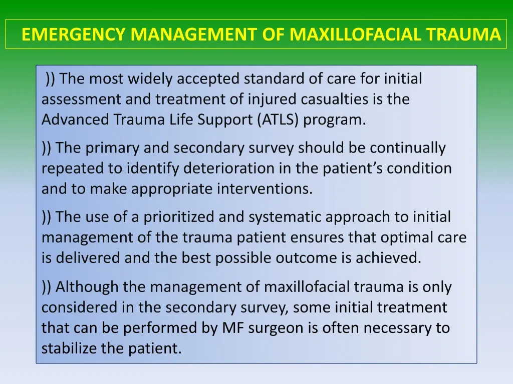
EMERGENCY MANAGEMENT OF MAXILLOFACIAL TRAUMA. )) The most widely accepted standard of care for initial assessment and treatment of injured casualties is the Advanced Trauma Life Support (ATLS) program.

E N D
EMERGENCY MANAGEMENT OF MAXILLOFACIAL TRAUMA )) The most widely accepted standard of care for initial assessment and treatment of injured casualties is the Advanced Trauma Life Support (ATLS) program. )) The primary and secondary survey should be continually repeated to identify deterioration in the patient’s condition and to make appropriate interventions. )) The use of a prioritized and systematic approach to initial management of the trauma patient ensures that optimal care is delivered and the best possible outcome is achieved. )) Although the management of maxillofacial trauma is only considered in the secondary survey, some initial treatment that can be performed by MF surgeon is often necessary to stabilize the patient.
The initial assessment should not concentrate on the most obvious injury but involve a rapid survey of the vital functions to allow management priorities to be established. The primary survey involves: A - Airway maintenance with cervical spine control B - Breathing and ventilation C - Circulation with haemorrhasecontrol D - Disability: neurological status E - Exposure: complete examination of the patient
During the primary survey, resuscitation is performed and afterwards shock management is initiated with fluid replacement whilst the vital signs are monitored for any deterioration in the status of the patient. Once the patient is stabilized a secondary survey is carried out to ensure that all traumatic injuries are evaluated, after which definitive care can be delivered.
A Airway assessment Signs of airway obstruction including stridor, gurgling, agitation, and hoarseness should be quickly assessed. The physician should evaluate for possible facial, mandibular, or tracheal or laryngeal fractures, which may compromise the airway and eventually lead to obstruction. The presence of blood, vomit, fractured teeth, or other debris in the oral cavity is concerning for potential airway compromise and should be monitored closely.
Trauma to the maxillofacial region can cause airway compromise because of hemorrhage, tissue swelling, and fractures leading to loss of facial architecture. Midfaceinjuries can compromise the nasopharynx and oropharynx as a result of fractures and dislocations. Severely comminuted or bilateral mandibularfractures may cause airway obstruction because of collapse of the glottic structures on the posterior pharynx. Dentoalveolarfractures, in addition to being associated with hemorrhage, can be problematic if teeth are dislodged because these can be easily aspirated. Therefore, all teeth should be accounted for to ensure none have been aspirated.
Airway management Control of the airway is an absolute priority. Initial management of the patient with airway compromise who requires intubation consists of a chin-lift or jaw-thrust maneuver, which is maintained until intubation is achieved. The tongue or palate is drawn forward and the mouth and pharynx sucked or wiped clear of debris to re-establish the airway. To keep the tongue forward, a suture may be passed through it, or the patient turned on his side, with control of the cervical spine, to allow saliva and blood to drain out of the mouth. Where these measures are ineffective intubation may be necessary
Indications for intubation of the trauma patient include airway obstruction; shock; altered mental status (Glasgow Coma Scale [GCS] ≤ 8); and occasionally combativeness requiring sedation for evaluation. Oropharyngealairways can serve as helpful adjuncts, but these cannot be used in conscious patients because of potential gagging, vomiting, and aspiration. Nasopharyhgeal airways are more tolerable in the awake patient and may transiently aid in maintaining airway patency.
When a decision has been made to initiate a definitive airway, orotracheal intubation is typically performed. In the urgent setting where orotracheal intubation is unsuccessful, prompt transition to a surgical airway (cricothyroidotomy or tracheostomy) is recommended.
B Breathing and ventilation Assessment of breathing and ventilation includes inspection, palpation, and auscultation of the neck, thoracic region, and upper abdomen and back. Injuries that can be identified during the primary survey and may restrict adequate ventilation include tension pneumothorax; flail chest (three or more consecutive ribs fractured in two places) with underlying pulmonary contusions; open pneumothorax; and massive hemothorax. Treatment is tubethoracostomyby placing a chest tube through the 2ndintercostal space for decompression.
C Circulation with hemorrhage control Shock is defined as inadequate tissue perfusion. The most common cause of shock in the injured patient is hemorrhagic in nature. Clinical signs of shock include tachycardia; dyspnea; cool and clammy skin; mental status changes; decreased pulse pressure; and in more severe cases, hypotension. Estimations of overall blood loss using vital signs has been suggested by ATLS to assist in determining optimal resuscitation strategies for patients in shock. As the severity of shock increases, recommendations for fluid replacement change from crystalloids to packed red blood cells (PRBC) and fresh frozen plasma (FFP).
Management of hemorrhage Pressure pack, clamping by hemostats, ligatures, ligation of external carotid artery, Posture, Electrocoagulation. Reduction and fixation will often arrest haemorrhage because movement disturbs the natural arrest. The pulse and blood pressure should be monitored to ensure that the patient does not become shocked, and appropriate fluid replacement should be started where necessary.
D Disability (neurologic status) The neurologic component of the primary survey quickly assesses the patient’s level of consciousness, pupillary size and reaction, and spinal cord injury level. The level of consciousness is determined using the GCS, which is composed of three criteria: (1) eye, (2) verbal, and (3) motor assessments.
GCS Scores of 14–15 are mild, 9–13 moderate, and 3–8 severe.
E Exposure and environmental control Patients should be completely exposed during the primary survey to fully examine and identify all injuries. However, prolonged exposure places the patient at risk for hypothermia, and therefore the examination should be completed as quickly as possible and the patient covered with warm blankets.
SECONDARY SURVEY Maxillofacial injuries are not typically life-threatening, so evaluation of injuries that do not involve airway obstruction or significant bleeding is delayed until the secondary survey.
History and Physical Examination After the patient has been initially stabilized, as complete a history as possible should be obtained (from the patient or his accompanies). Evaluation of the facial area should be performed in an organized and sequential fashion. The face and cranium should be carefully inspected for evidence of trauma, including lacerations, abrasions, contusions, areas of edema or hematoma formation, and possible contour defects. Areas of ecchymosis should be carefully evaluated. Periorbitalecchymosis, especially with subconjunctival hemorrhage, is often indicative of orbital rim or zygomatic complex fractures. Bruises behind the ear, or the Battle sign, suggest a basilar skull fracture. Ecchymosis in the floor of the mouth usually indicates an anterior mandibular fracture.
A complete intraoral examination should be performed at this point to assess for any lacerations, floor of mouth hematomas, or malocclusion, which may be indicators of alveolar, maxillary, or mandibular fractures.
Having carried out a thorough inspection of the mouth and face, the facial skeleton should be palpated in the same systematic manner, paying particular regard to:
Stand behind and above the patient when assessing facial asymmetry, particularly in relation to suspected zygomaticfractures. In this position place an index finger on the maximum convexity of the zygomaon both sides equidistant from the tip of the nose. Then compare the overlap of the index fingers with the supraorbitalridges.
The mandible should be carefully evaluated by extraorallypalpating all areas of the inferior and lateral borders and the TMJ, paying particular attention to areas of point tenderness. Bimanual palpation of the suspected fracture area should be performed by placing firm pressure over the mandible posterior and anterior to the fracture area in an attempt to manipulate and elicit mobility in this area. The occlusion should be re-examined after this maneuver. Mobility of the teeth in the area of a possible fracture should also be noted.
Mobility of the middle third of the face, such as that brought about by Le Fort I, Le Fort II and Le Fort III pattern fractures is best assessed by placing the patient’s head securely against a head rest, grasping the upper teeth and alveolus and moving them gently, but purposefully, laterally, superiorly and anteriorly. A ‘cracked cup’ sound when the upper teeth are percussed can be diagnostic of a Le Fort pattern fracture.
Diplopia (double vision) is most often caused by haemorrhage or oedema in or adjacent to extraocular muscles, but can be caused by mechanical tethering of muscle attachments or by injuries to the third, fourth or sixth cranial nerves. Visual acuity or pupillary changes may suggest intracranial (cranial nerve II or III dysfunction) or direct orbital trauma. Restricted eye ball movement in any direction of gaze may indicate orbital fracture and entrapment of muscles. Posterior displacement of the globe gives rise to enophthalmos due to a ‘blowout’ fracture of the orbital floor or medial wall.
An evaluation of the nose and paranasal structures includes measurement of the intercanthal distance between the innermost portions of the left and right medial canthus. Frequently, naso-orbital ethmoidinjuries cause spreading of the nasal bones and displacement of the medial canthal ligaments, resulting in traumatic telecanthus (widening of the medial intercanthaldistance).
Radiographic assessment Radiographic examination forms the basis of special investigations of maxillofacial injury and should be specific to the areas of concern. Radiographs may be necessary to inform treatment decisions, for example, about where bone plates should be applied. They also commonly reveal injuries which may not need treatment, for example, undisplaced or minimally displaced fractures, medial blow-out fractures of the orbit and comminution of the midface.
CT is the most commonly used radiographic technique for evaluation of midface trauma. The ability to evaluate fractures in several planes of space and to visualize the entire skull, midfaceand mandible with three-dimensional reconstruction provides invaluable information for diagnosing and treating complex facial trauma To be continued…