Download
1 / 77
770 likes | 912 Views
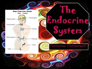
The Endocrine System. Hypophyseal-Pituitary Axis. Site of Neural – Hormonal interaction Sets temporal release of hormones Responsible for stress reaction of hormones. The Hypothalamus: Located in the brain, this region controls most endocrine secretions

E N D
Hypophyseal-Pituitary Axis • Site of Neural – Hormonal interaction • Sets temporal release of hormones • Responsible for stress reaction of hormones
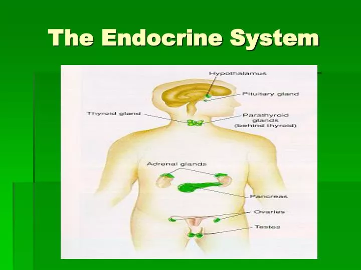
The Hypothalamus: Located in the brain, this region controls most endocrine secretions Mainly regulatory hormones are released here. Most control the pituitary gland The Pituitary Gland Descending from the hypothalamus, this gland has two halves: anterior & posterior The anterior half secretes mainly regulatory hormones The posterior half secretes hormones, but manufactures none The Hypothalamus & the Pituitary Gland-- Master Endocrine Glands!
Hormones secreted by the Hypothalamus & Anterior Pituitary Gland Hypothalamus Anterior Pituitary GHRH (GH-releasing) GH (growth hormone) SS (somatostatin, GH-inhib) “ CRH (corticotropin-rel) ACTH (adrenocorticotropic) GnRH (gonadotropin-rel) LH (luteinizing hormone) “ FSH (follicle-stimulating) PRH (PRL-releasing) PRL (prolactin) PIH (PRL rel-inhibiting) “ TRH (thyrotropin-rel) TSH (thyroid stimulating)
Growth Hormone: stimulates cells to grow and divide increases amino acid transport rate and protein synthesis increases fat metabolism Typically, GH is secreted during sleep. GH secretion increases when malnourished GH influences bone growth via somatomedin: GH in blood GH arrives in liver liver secretes somatomedin cartilage divides bones grow! What do these anterior pituitary hormones do?
Problems with GH • Too much GH in children leads to gigantism • Too much GH in adults leads to acromegaly • Too little GH in children leads to dwarfism
ACTH: works on the cortex of the adrenal gland, influencing the release of cortisol stress can increase CRH secretion which will increase ACTH secretion negative feedback when adrenal cortex hormones in blood decrease CRH secretion LH & FSH: LH in females and in males leads to sex hormone secretion FSH in females causes growth and development of egg cell-containing follicles in the ovary, and causes estrogen secretion FSH in males instigates sperm production both hormones are regulated by GnRH, which is not significant in concentration until puberty Other Anterior Pituitary Hormone Functions
PRL: In females, PRL promotes lactation In males, PRL decreases LH secretion (note that too much PRL would then decrease androgen levels and cause sterility) Controlled by both PRH and PIH TSH: works on thyroid gland to either cause or inhibit its secretion of hormones works on thyroid gland to affect its growth (too much TSH leads to a goiter) negative feedback via thyroid hormones in blood stress or cold temperatures can change TSH secretion More Anterior Pituitary Hormone Functions
The Posterior Pituitary Lobe No hormones are made here. They are made in the hypothalamus and just released here. Two peptide hormones are released from the posterior pituitary lobe (the neurohypophysis): • ADH (antidiuretic hormone or vasopressin) • OT (oxytocin)
ADH: as an “antidiuretic,” ADH decreases urine formation by having kidneys conserve water also can contract smooth muscle cells, as found in blood vessels-- this causes an increase in blood pressure ADH release triggered by osmoreceptors and inhibited by stretch receptors in blood vessels OT: In females, contracts the uterine wall smooth muscles In females, helps to eject milk when lactating No known function in males, although in both males and females, OT can have some antidiuretic effects Function of Posterior Pituitary Lobe Hormones
Hypothalamus & Anterior Pituitary Hypothalamus Anterior Pituitary GHRH (GH-releasing) GH (growth hormone) SS (somatostatin, GH-inhib) “ CRH (corticotropin-rel) ACTH (adrenocorticotropic) GnRH (gonadotropin-rel) LH (luteinizing hormone) “ FSH (follicle-stimulating) PRH (PRL-releasing) PRL (prolactin) PIH (PRL rel-inhibiting) “ TRH (thyrotropin-rel) TSH (thyroid stimulating)
HPA Basics • Hypophysis • Third Ventricle • GRH, TRH, CRH, GnRH, Dopamine, Somatostatin • Neurohypophysis • Derived from Hypophysis • ADH, Oxytocin • Adenohypophysis • Derived from Rathke’s pouch • ACTH, LH, FSH, TSH, GH, PRL
Pituitary Diseases • Primary Tumors • Adenomas • Craniopharyngioma • Metastasis • Empty Sella • Surgical, post-Sheehand’s • Hemorrhage • Sheehand’s syndrome • Hyperfunction • Prolactin • Insufficiency
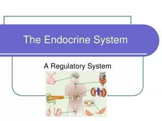
The Endocrine Glands and Their Hormones • Pituitary Gland • A marble-sized gland at the base of the brain • Controlled by the hypothalamus or other neural mechanisms and therefore the middle man. • Posterior Lobe • Antidiuretic hormone: responsible for fluid retention • Oxytocin: contraction of the uterus
The Endocrine Glands and their Hormones • Pituitary Gland • Exercise appears to be a strong stimulant to the hypothalamus for the release of all anterior pituitary hormones • Anterior Lobe • Adrenocorticotropin • Growth hormone * • Thyropin • Follicle-stimulating hormone • Luteinizing hormone * • Prolactin
The Endocrine Glands and Their Hormones • Thyroid Gland • Located along the midline of the neck • Secretes two nonsteroid hormones • Triiodothyronine (T3) • Thyroxine (T4) • Regulates metabolism • increases protein synthesis • promotes glycolysis, gluconeogenesis, glucose uptake • Calcitonin: calcium metabolism
Thyroid • Largest Endocrine organ in the body • Involved in production, storage, and release of thyroid hormone • Function influenced by • Central axis (TRH) • Pituitary function (TSH) • Comorbid diseases (Cirrhosis, Graves, etc.) • Environmental factors (iodine intake)
The Thyroid Gland Structure: This bilobed gland contains many follicles. A follicle is a group of cells encircling a lumen. The lumen contains material called colloid (a glycoprotein) within it. As hormones are produced by the cells, the hormones are either released into the colloid or directly into the blood. There are also extrafollicular hormone-secreting cells, called C cells. These are found between lumina. Hormones Produced: • Thyroxine (T4) made in follicle • Triiodotyronine (T3) made in follicle • Calcitonin made by C cells
T3 and T4: Function: metabolism regulation (break down carbohydrates and fats, synthesize proteins) Can only be made by follicular cells when iodides are available Somewhat hydrophobic and get carried by proteins in the blood. Controlled by anterior pituitary lobe TSH T3 more effective, T4 more abundant Calcitonin: Function: decrease blood calcium levels and blood phosphate levels (by helping them get deposited in bone, and by stimulating excretion of them by kidneys) Controlled by blood calcium levels and digestive chemicals About the Thyroid Hormones...
Problems with the Thyroid Gland Hyperthyroidism: • high metabolic rate, hyperactivity, sensitivity to heat, protruding eyes • Grave’s disease: when hyperthyroidism is due to an autoimmune problem (TSH is mimicked by autoantibodies) Hypothyroidism: • in the adult: low metabolic rate, sensitivity to cold, sluggishness • in an infant: cretinism-- stunted growth, mental retardation, abnormal bone formation • Hashimoto’s disease: when hypothyroidism is due to an autoimmune problem (autoantibodies attack and destroy follicular cells) • goiter: no T3 and T4 can be made because not enough iodides were ingested.
Thyroid (cont) • Regulates basal metabolic rate • Improves cardiac contractility • Increases the gain of catecholamines • Increases bowel motility • Increases speed of muscle contraction • Decreases cholesterol (LDL) • Required for proper fetal neural growth
Thyroid Physiology • Uptake of Iodine by thyroid • Coupling of Iodine to Thyroglobulin • Storage of MIT / DIT in follicular space • Re-absorption of MIT / DIT • Formation of T3, T4 from MIT / DIT • Release of T3, T4 into serum • Breakdown of T3, T4 with release of Iodine
Iodine uptake • Na+/I- symport protein controls serum I- uptake • Based on Na+/K+ antiport potential • Stimulated by TSH • Inhibited by Perchlorate
Secretion of Thyroid Hormone • Stimulated by TSH • Endocytosis of colloid on apical membrane • Coupling of MIT & DIT residues • Catalyzed by TPO • MIT + DIT = T3 • DIT + DIT = T4 • Hydrolysis of Thyroglobulin • Release of T3, T4 • Release inhibited by Lithium
Thyroid Hormone • Majority of circulating hormone is T4 • 98.5% T4 • 1.5% T3 • Total Hormone load is influenced by serum binding proteins (TBP, Albumin, ??) • Thyroid Binding Globulin 70% • Albumin 15% • Transthyretin 10% • Regulation is based on the free component of thyroid hormone
Hormone Binding Factors • Increased TBG • High estrogen states (pregnancy, OCP, HRT, Tamoxifen) • Liver disease (early) • Decreased TBG • Androgens or anabolic steroids • Liver disease (late) • Binding Site Competition • NSAID’s • Furosemide IV • Anticonvulsants (Phenytoin, Carbamazepine)
Hormone Degredation • T4 is converted to T3 (active) by 5’ deiodinase • T4 can be converted to rT3 (inactive) by 5 deiodinase • T3 is converted to rT2 (inactive)by 5 deiodinase • rT3 is inactive but measured by serum tests
TRH • Produced by Hypothalamus • Release is pulsatile, circadian • Downregulated by T4, T3 • Travels through portal venous system to adenohypophysis • Stimulates TSH formation
TSH • Produced by Adenohypophysis Thyrotrophs • Upregulated by TRH • Downregulated by T4, T3 • Travels through portal venous system to cavernous sinus, body. • Stimulates several processes • Iodine uptake • Colloid endocytosis • Growth of thyroid gland
Iodine states • Normal Thyroid • Inactive Thyroid • Hyperactive Thyroid
Hypothyroid • Symptoms – fatigability, coldness, weight gain, constipation, low voice • Signs – Cool skin, dry skin, swelling of face/hands/legs, slow reflexes, myxedema • Newborn – Retardation, short stature, swelling of face/hands, possible deafness • Types of Hypothyroidism • Primary – Thyroid gland failure • Secondary – Pituitary failure • Tertiary – Hypothalamic failure • Peripheral resistance
Hypothyroid • Cause is determined by geography • Diagnosis • Low FT4, High TSH (Primary, check for antibodies) • Low FT4, Low TSH (Secondary or Tertiary, TRH stimulation test, MRI) • Treatment • Levothyroxine (T4) due to longer half life • Treatment prevents bone loss, cardiomyopathy, myxedema
Goiter • Endemic goiter • Caused by dietary deficiency of Iodide • Increased TSH stimulates gland growth • Also results in cretinism • Goiter in developed countries • Hashimoto’s thryoiditis • Subacute thyroiditis • Other causes • Excess Iodide (Amiodarone, Kelp, Lithium) • Adenoma, Malignancy • Genetic / Familial hormone synthesis defects
Hyperthyroid • Symptoms – Palpitations, nervousness, fatigue, diarrhea, sweating, heat intolerance • Signs – Thyroid enlargement (?), tremor • Lab workup • TSH • FT4 • RAIU • Other Labs • Anti-TSH-R Ab, Anti-TPO Ab, Anti-TBG Ab • FT3 • FNA • MRI, US
Hyperthyroid • Common Causes • *Graves • Adenoma • Multinodular Goiter • *Subacute Thyroiditis • *Hashimoto’s Thyroiditis • Rare Causes • Thyrotoxicosis factitia, struma ovarii, thyroid metastasis, TSH-secreting tumor, hamburger
The Endocrine Glands • Parathyroid Glands • Secretes parathyroid hormone • regulates plasma calcium (osteoclast activity) • regulates phosphate levels
Calcium Regulation Parathyroid
Calcium • Required for muscle contraction, intracellular messenger systems, cardiac repolarization. • Exists in free and bound states • Albumin (40% total calcium) • Phosphate and Citrate (10% total calcium) • Concentration of iCa++ mediated by • Parathyroid gland • Parafollicular C cells • Kidney • Bone
Parathyroid Gland • This gland only secretes one hormone: Parathyroid Hormone (or PTH) • PTH function (we began learning this when we studied bone): • increases blood calcium (Ca2+) levels and decreases blood phosphate (PO42-) levels
PTH function (continued) • How does PTH work? • PTH causes Ca2+ & PO42- to be released from bone into blood (by increasing osteoclast activity) • PTH causes the kidneys to remove PO42- ions from the urine • PTH increases vitamin D production, so that you absorb more Ca2+ during digestion • PTH is regulated by blood calcium levels-- not by other glands!