Download
1 / 17
180 likes | 453 Views
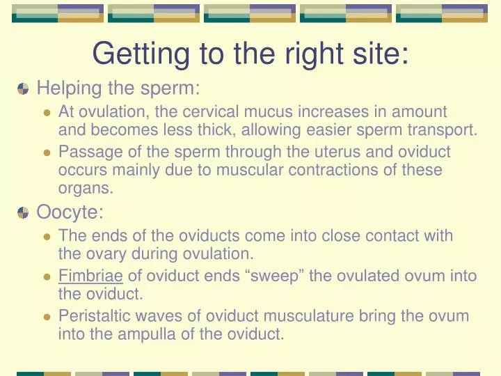
Getting to the right site:. Helping the sperm: At ovulation, the cervical mucus increases in amount and becomes less thick, allowing easier sperm transport. Passage of the sperm through the uterus and oviduct occurs mainly due to muscular contractions of these organs. Oocyte:

E N D
Getting to the right site: • Helping the sperm: • At ovulation, the cervical mucus increases in amount and becomes less thick, allowing easier sperm transport. • Passage of the sperm through the uterus and oviduct occurs mainly due to muscular contractions of these organs. • Oocyte: • The ends of the oviducts come into close contact with the ovary during ovulation. • Fimbriae of oviduct ends “sweep” the ovulated ovum into the oviduct. • Peristaltic waves of oviduct musculature bring the ovum into the ampulla of the oviduct.
The OviductH & E × 10M = muscleBL = broad ligamentS = serosa with vascular supporting tissue From Wheater’s Functional Histology, 4th ed., 2000.
Morphology of the Oviduct: Fallopian tube S = smooth muscle H & E × 150 E = ciliated epithelium Fallopian tube Azan × 320 From Wheater’s Functional Histology, 4th ed., 2000.
Capacitation: readying the sperm • Sperms cannot fertilize oocytes when they are newly ejaculated. • The process of capacitation takes 5-7 hours. • Capacitated sperms are more active. • Location: capacitation occurs in the uterus and oviducts and is facilitated by substances of the female genital tract. • The acrosomal reaction cannot occur until capacitation has occurred.
Stage 1 of fertilization: • The acrosome reaction must be completed before the sperm can fuse with the secondary oocyte • Occurs when sperms come into contact with the corona radiata of the oocyte • Perforations develop in the acrosome • Point fusions of the sperm plasma membrane and the external acrosomal membrane occur • The acrosome reaction is associated with the release of acrosome enzymes that facilitate fertilization • Passage of sperm through the corona radiata depends on enzyme action: • hyaluronidase released from sperm acrosome • Tubal mucosal enzymes • Flagella action also aids corona radiata penetration
Ovum and sperms: (In vitro) From this photograph, it should be clear that the heads of human sperm are less than 1/20 the diameter of human eggs. Arrows point to sperm heads Advanced Fertility Center of Chicagohttp://www.advancedfertility.com/ The surfaces of unfertilized eggs are usually smooth in appearance. The mottled look of this egg is not normally seen, but apparently all the ova from this woman had this appearance.
Stage 2 of fertilization: • Penetration of the zona pellucida around the oocyte: • Acrosomal enzymes: esterases, acrosin, and neuraminidase cause lysis of the zona pellucida • Once sperm penetrates zona pellucida, the zona reaction occurs: • This reaction makes the zona pellucida impermeable to other sperms. • When more than one sperm manages to enter the ovum (dispermy = 2; triploidy = 3), the fetus nearly always aborts.
Stages 3 & 4 of fertilization: • Fusion of plasma membranes of oocyte and sperm • Head and tail of a sperm enter the cytoplasm of the oocyte, but the sperm plasma membrane remains behind. • 2nd meiotic division of oocyte is completed • The secondary oocyte was previously arrested in metaphase of the 2nd meiotic division, and now forms the mature ovum and another polar body.
Stage 5 of fertilization: • Formation of male and female pronuclei: • Chromosomal material of the sperm decondensates and enlarges • Chromosomal material of the ovum decondensates following the completion of meiosis • At this stage, the male and female pronuclei are indistinguishable. • As they grow, the pronuclei replicate their DNA still 1N (haploid)- 23 chromosomes, each in chromatid pairs
Fusion of the pronuclei: (in vitro) • The male and female pronuclei are indistinguishable from one another. • The second polar body can be seen (arrow). • The plasma membranes of the two pronuclei are dissolving and one diploid nucleus will remain. Advanced Fertility Center of Chicagohttp://www.advancedfertility.com/
Stage 6 of fertilization: • Membranes of the pronuclei break down, chromosomes condense and arrange themselves for mitotic cell division • On membrane dissolution, there is 1 cell with 46 chromosomes = diploid (2N) • The first cleavage follows shortly, leaving 2 cells, each with 46 chromosomes. • Mitosis in the new zygote uses centrioles derived from the sperm. The oocyte has no centrioles.
Fertilization facts: • Completed within 24 hours of ovulation • Approximately 400 to 600 MILLION sperms are deposited at cervical opening during ejaculation. • Some sperm are held up by the folds of the cervix and are gradually released into the cervical canal; this gradual release increases the chances of fertilization. • Most human sperms do not survive longer than 48 hours in the female genital tract. • Only about 200 sperms reach the fertilization site; most degenerate and are absorbed by the female genital tract.
The results of fertilization: • Stimulates the secondary oocyte to complete meiosis. • Restores the normal diploid number of chromosomes (46). • Results in variation of human species as maternal and paternal chromosomes intermingle. • The embryo contains only maternal mitochondria because the sperm mitochondria are dispersed into the egg cytoplasm and discarded. • Determines the sex of the embryo. • The sex chromosome (Y or X) carried by the successful sperm determines embryonic sex.
Twins: still 1 sperm per egg • Monozygotic (monoovular): • A fertilized, single egg splits into two developing zygotes at a very early stage. • Identical twins; same sex. • Dizygotic (polyovular): • Result from the fertilization by two sperm of two separate ova that have reached maturation at the same time. • Not identical twins; can be different sexes • Incidence increases with age of the mother
How can fertilization go awry? • Too many sperm = dispermy or triploidy • Leads to spontaneous abortion in most cases. • Infertility • Bad timing: • The sperm can only survive 48 hours within the female genital tract. • In vitro studies show the ovulated egg cannot be fertilized after 24 hours.
Triploidy (in vitro) There are 3 pronuclei within this one zygote. In the laboratory, such embryos are discarded. In vivo, such embryos almost always abort spontaneously. Advanced Fertility Center of Chicagohttp://www.advancedfertility.com/
A little more on sex determination: • Not so clear-cut as X and Y: • The presence of other genes, located on the X, Y and other chromosomes, influence the determination of embryonic sex. • The SRY gene: • Normally present on the Y chromosome • Induces the sexless embryonic gonad to develop into a testis • Can sometimes be present on the X chromosome, leading a genetic female (XX) to develop testis • 1990: a fertilized mouse embryo with the XX configuration was injected with SRY and developed as a male both anatomically and behaviorally. • (Andrew Sinclair, Center for Hormone Research, University of Melbourne)