Download
1 / 34
450 likes | 985 Views
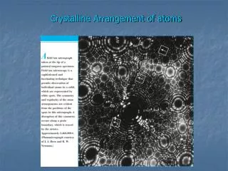
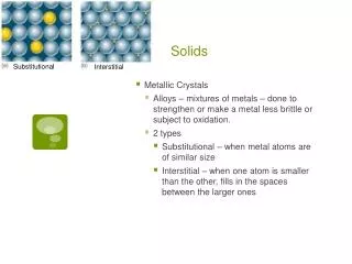
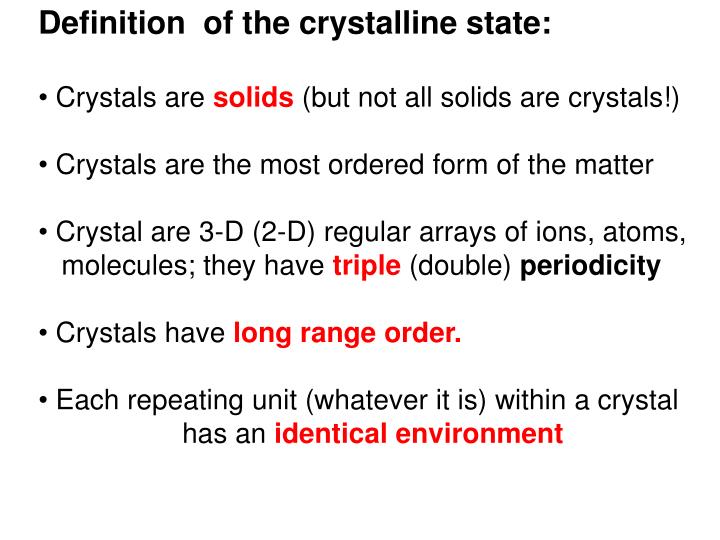
Definition of the crystalline state: Crystals are solids (but not all solids are crystals!) Crystals are the most ordered form of the matter Crystal are 3-D (2-D) regular arrays of ions, atoms, molecules; they have triple (double) periodicity Crystals have long range order.

E N D
Definition of the crystalline state: • Crystals are solids (but not all solids are crystals!) • Crystals are the most ordered form of the matter • Crystal are 3-D (2-D) regular arrays of ions, atoms, • molecules; they have triple (double) periodicity • Crystals have long range order. • Each repeating unit (whatever it is) within a crystal • has an identical environment
X-ray Diffraction is the essence of the X-ray crystal structure analysis (XRA) • The main aim of XRA is the determination of 3-D structure of the chemical • entity = structural motive which is forming the crystal that is being • repeated periodically in the whole volume of the crystal • Crystal forming chemical entities = motives: metals, ions, atoms (e.g. diamond), • organic compounds, peptides, proteins, lipids, oligosaccharides, DNA, RNA etc.
X-ray analysis is the most accurate method to determine: - structure of the crystal hence structure of the crystal motif e.g. molecule: - bond distances and angles(C-C 1.542(2) Å, C-C-N 123.72(12)o) - conformation of the compound - absolute configuration of the compound/atom X-ray crystal analysis is the best source of the above data: they are the key components of structural databases for further chemical (e.g. quantum) and physical calculations Co(III)(Ph4porphyrin) (Cl) Rh(en)2Cl2+ trans Mn(H2O)62+
What are the components of waves? |F| A |F| B wavelength | F | = amplitude phase
How to obtain the structure of the motif (compound) from its crystal structure? The foundation of recognition/”visibility” of all structures: Scattering and Diffraction All objects – irrelevant of their size – scatter radiation which is shine on them. http://www.acoustics.salford.ac.uk/feschools/waves/super.htm#phase http://www.numathics.com/arens/scattering/Scattering.html
Difference between scattering and diffraction: - scattering: spherical - diffraction: more directional as it results from interference of scattering from many centres, - (or it results from interference of incoming = = source wave with new, scattered waves.)
How we can retrieve the information about the scatteringobjects? Because we can focus back the scattered/diffracted waves again. Theimage of the object Lenses: focussing Scattering/Diffraction Theobject
Why can we focus back the image of the scattering/diffracting object? > • Because light travels with different media with different speed: - different media have different refractive index n • n is a measure how much is the speed of light (or other waves such as sound waves) is reduced inside the medium > nG nA Air nAc nG Glass nG = 1.5 Here
What should be the relationship between effective scattering and the size of the object ? Web examples http://www.acoustics.salford.ac.uk/feschools/waves/super.htm#phase • The power of scattering/diffraction by an object is: • directly proportional to the similarity between the wavelength of the incident radiation and the size of the scattering object • Larger object – larger waves needed for an effective scattering • Smaller objects – smaller wave needed for an effective scattering
Typical C-C bond:Å, so The most useful radiation to study crystal Structure has to have in the rangeÅ Hence: X-ray radiation in X-ray analysis! 1.54 ~1-2 of radiation 3.7 pm 400 – 700 nm Resolution 2 nm 10 nm 200nm 1 meter = 102 cm = 103 mm = 106 mm = 109 nm =1010Å
wave = | F| x = F Intensity of the wave: I ~ F 2 If we know I then: I = √F 2 = |F| Lenses are focussing back all information that is contained in the scattered or diffracted waves: - amplitudes|F | (intensities I) - phases so they can produce back the image of the scattering/diffracting object
What?!! NO LENSES!!!! Usually Monochromatic: one, well defined In X-ray Diffraction we do not have lenses which could focus diffracted rays back to the crystal structure, n=1!. We can only register: - directions of the diffracted X-rays and their - intensitiesI hence |F|amplitudesonly of the diffracted rays: !their phases are missing! = phase problem
Information directly available from an X-ray single crystal diffraction experiment: 1. IntensitiesIof diffracted X-rays therefore|F | - amplitudes of diffracted X-rays 2. Directions of the diffracted X-rays The phases must be reconstructed in rather complex/difficult experimental and computing methods: phase problem = phase solution methods
The key-feature of XRD and XRA is the interaction between • the crystal and the incoming X-ray radiation • (l in range off 0.8 – 2 Å). • X-rays in the crystal are: • scattered by the electrons: • - Thomson scattering: the electron oscillates in the • electric field of the incoming X-ray beam and an • oscillating electric charge radiates electromagnetic waves • - this is elastic and coherent scattering: • frequencies and wavelengths of the incoming X-rays • and scattered-diffracted X-rays are the same/unchanged • this scattering is becoming very discreet in terms of directions • some scattered X-ray waves are reinforced, some weakened • as we are dealing here with the diffraction - REFLECTIONS • which is amplified by millions copies of the same atoms (electrons!) • in the same positions in the crystal space due to • crystal periodic, repetitive (in 3-D) unique character • But why not to measure scattering from one molecule and determine its • structure this way?…..
We cannot measure (yet) the X-ray scattering produced by single chemical entity (organic molecule): it is too weak. • We use crystals as 3-D amplifiers of scattering • coming from single crystal motif. • X-ray Diffraction is well welcomed “side effect” of this process due to amplifying or cancelling effect • of scattered radiation emitted by electrons • There are also other types of interactions of X-rays with electrons: e.g. excitations. • These type of high energy phenomena would damage the • single molecule almost immediately. • In crystal there are thousands of molecules – some of them survive long enough to give a measurable radiation. • .
Crystalstructure = CrystalLattice + motifweb: http://marie.epfl.ch/x-ray/rlattice/ = + The 3-D periodicity of the crystal can be simplified and represented by an abstract crystal lattice.
Crystal lattice is described by three translations: a, b, c • They can not be just any translations: they have to reproduce all crystal motives (lattice points) if applied to any single lattice point ? It is NOT the unit cell ? b a It is the right unit cell Two atom compound: X
They determine the unit cell, which has to be: • a lattice ‘building block’,which edges correspond toa, b, c • itshould give the whole crystal lattice if moved by a, b, c • it has to be of theright handed system • it has to have thesmallest possible volume • it has posses thehighest possible symmetry characteristic for the lattice (this is why some unit cells are not primitive)
z c b y a x The unit cell in three dimensions. The unit cell is defined by three vectors a, b, and c, and three angles , , . Unit cells are defined in terms of the lengths of the three vectors and the three angles between them. For example, a = 94.2 Å, b = 72.6 Å, c = 30.1 Å, = 90.0°, = 102.1°, = 90.0° a = 8.32 Å, b = 15.23 Å, c = 9.28 Å, a = 90.0°, b = 90.0°, g = 90.0°
Content of the Unit cell c Size and the arrangement (symmetry) of the unit cell Crystal structure Motif = molecule, atoms To get the structure of the motive we have to: get the information about the unit cell size and its arrangement
z (u,v,w) y c b a x In the crystal lattice we can distinguish: - lattice points - lattice directions - lattice planes Co-ordinates of the lattice points are given in the fractions u,v,w of the a,b,c lattice translations
Crystal Lattice directions symbol: examples: [uvw] [100][010] [001] : z : c b a c y x b a
Crystal planes = inter-plane spacing measured at 90o to the planes • The planes are “imaginary” • All planes in a set of planes are identical - equivalent • The perpendicular distance between pairs of adjacent planes is called d: interplanar spacing • Need to label planes to be able to identify them…………
Find intercepts on a, b, c:1/2, 1, 0 (1 1/3 0) Take reciprocals 2, 1, 0 (1 3 0) (1 3 0) plane plane (2 1 0) Miller index (hkl) (h kl) (h k l) General label is (h k l)which intersects at a/h, b/k, c/l (hkl) is the MILLER INDEX of that plane (round brackets, no commas).
z 1c O y x 1b 1a z (222) 1c y O x 1b 1a (111) Standard triangle
Plane perpendicular to x cuts at 1, , (1 0 0) plane c b (0 1 0) plane a (0 0 1) plane c c NB an index 0 means that the plane is parallel to that axis b b a a
Planes - conclusions • Miller indices define the orientationof the plane within the unit cell • The Miller Index defines a set of planesparallel to one another (remember the unit cell is a subset of the “infinite” crystal • All possible sets of planes in a particular lattice may be described by (hkl) values • Any of these sets of planes may contain scattering electrons (atoms) (or be close to): this is crucial for scattering and diffraction. • Distance between planes is given by dhkl • Reciprocal dependence between (hkl) and dhkl: • Larger (hkl) values (finely spaced planes) then smaller dhkl.
Diffraction – on the optical grating Web example! Path difference XY between diffracted beams 1 and 2: sin = XY/a XY = a sin For 1 and 2 to be in phase and give constructive interference, XY = , 2, 3, 4…..n so a sin = n where n is the order of diffraction It is so-called grating relationship where a = is the distance between scattering centres
Principles of BRAGG X-ray diffraction experiment: diffracted X-rays Crystal Incoming X-ray Non-diffracted X-rays (97%) detector
Diffraction – on the crystal lattice“grating” Incident X-ray radiation “Reflected” radiation 1 (ca. 3%) 2 dhkl Set of crystal planes (h k l) X Z Y dhkl dhkl Transmitted radiation XY = YZ = dhklsin (ca. 97%!) Beam 2 lags beam 1 by XY + YZ = 2d sin So 2dhkl sin = n Bragg Law as inter-atomic distances are in the range of 0.5 - 2 Å so must be in the range of 0.5 - 2 Å X-rays, electrons, neutrons suitable
Difference between light and X-ray reflections + + + + + + + + - + - + - + - + Light: X-ray: • Reflection of the Light: • is not coherent (multi ) • does not depend on • is happening only on the surface • can be focused back • almost 100% of the incident light • is reflected • X-ray Diffraction/Reflection: • coherent (usually single ) • strictly depends on • is happening in 3-D volume of the crystal • can not be focused back • only about 3% of the incoming X-rays • is diffracted; ~97% goes • through the crystal unchanged