Download
1 / 68
750 likes | 1.06k Views
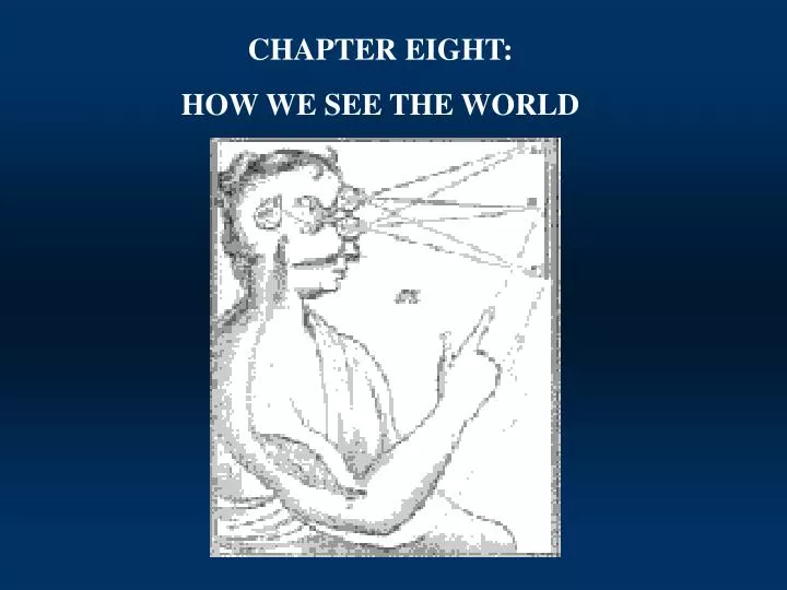
CHAPTER EIGHT: HOW WE SEE THE WORLD. We see the lines as different because we have been "taught" to use specific shapes and angles to tell us about size. Which of the lines shown below is longer?. THE NATURE OF SENSORY EXPERIENCE.

E N D
CHAPTER EIGHT: HOW WE SEE THE WORLD
We see the lines as different because we have been "taught" to use specific shapes and angles to tell us about size. Which of the lines shown below is longer? THE NATURE OF SENSORY EXPERIENCE • Vision and other sensory experiences of the world are not ‘genuine reproductions’ of the world’ rather, they are subjective interpretations created in the mind of the perceiver.
Electrical Fields Magnetic Fields THE NATURE OF SENSORY EXPERIENCE • Umwelt: species-specific perceptual world Perception is constructed differently among differing species.
Light: The Stimulus for Vision • We see light or electromagnetic energy which comes to us: • Directly from a light producing object (a light bulb) • Indirectly from a light source reflecting off an object • Light waves come in different lengths • The small range of ‘visible light’ is due to properties of visual receptors, not properties of the light itself. • Inter-species differences in light range detection
The Eye Cornea: Transparent tissue covering the front of the eye. Does not have any blood vessels; does have nerves. Iris: Circular band of muscles that controls the size of the pupil. The pigmentation of the iris gives "color" to the eye. Lens: Transparent tissue that bends light passing through the eye. To focus light, the lens can change shape by bending. Pupil: Hole in the center of the eye where light passes through. Retina: Layer of tissue on the back portion of the eye that contains cells responsive to light (photoreceptors) Sclera: Protect coating around the posterior five-sixths of the eyeball
The Eye • Light passes through the cornea, pupil, and lens on its way to the retina photoreceptors, which absorb and then convert it into electrical potentials that carry information to the brain. • The lens projects an inverted image onto the retina and the brain adjusts this inversion so we see the world in its correct orientation. • The cornea and lens bend or refract light rays as they enter the eye, in order to focus images on the retina. The eye can change the extent to which rays are bent and thus can focus images of objects by varying curvature of the lens. • Through the ciliary muscle • This ciliary muscle is smooth or involuntary muscle
The Photoreceptors • A sensory receptor is a specialized neuron that detects a specific physical stimulus. *Note: Do not confuse a sensory receptor with the protein molecules that function as neurotransmitter receptors in the membranes of neurons. • Most sensory receptors do not have axons. Their cell bodies synapse on dendrites or cell bodies of neurons.
The Photoreceptors • The photoreceptors process the light energy. • There are two types of photoreceptors: "rods" and "cones." • Rods are sensitive enough to respond to a single photon, but functioning together they are optimized for seeing in poor light. • Cones are optimized for responding to fine detail and color; they need a lot more light and work best in broad daylight. • Inside the human eye, there are eighteen times more rods than cones. These are arranged in such a way as to produce the best possible combination of night and day vision.
The Photoreceptors: located in the back of the retina When light hits the retina it it first processed in the photoreceptors located in the back of the retina. PHOTORECEPTORS BIPOLAR CELLS GANGION CELLS The ganglion cells’ axons join together to form the optic nerve, which exits through the back of the eye at the optic disk.
THE OPTIC DISK: YOUR BLIND SPOT close your right eye. With your left eye, look at the red circle. Slowly move your head closer to the image. At a certain distance, the blue line will not look broken!!
Sensation of whole spectrum of colors provided by exciting differently the 3 cone types The Photoreceptors: visual spectrum
The Photoreceptors: RODS • 120 million rods distributed over most of the retina except near the fovea • There are no rods at the very center of your visual field; we can’t see detail in poor illumination • Rods are connected in groups; there are far fewer optic nerves going to the brain than rods • Rod vision detects edges and motion very well • Rod pigment is bleached by light and is less effective in bright light; rods take about 20 - 30 minutes of ‘dark adaptation’ before they are most efficient
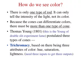
The Photoreceptors: CONES • 5 million cones • There is a concentration in fovea, region about 1.5 mm in diameter. Most acute vision limited to foveola, covering ~0.4 mm • No rods at all in central part of fovea • Color vision is provided by 3 types of cone with different colored light absorptions: red, green, and blue cones.
The Photoreceptors: PHOTOTRANSDUCTION • Rods and cones contain photopigments- chemicals that release energy when struck by light. • Photopigments consist of 11-cis-retinal (derivative of Vitamin A) which is bound to opsin (a protein). • Light converts 11-cis-retinal to all-trans-retinal, which ultimately activates 2nd messenger systems that work to close NA+ channels, hyperpolarizing the receptor. • More light = hyperpolarization.
The Photoreceptors: PHOTOTRANSDUCTION Photoreceptors input to retinal bipolar cells, which input to retinal ganglion cells. Photoreceptors and bipolar cells do not produce action potentials. They produce graded potentials. light Retinal ganglion cells produce action potentials.
The Photoreceptors: PHOTOTRANSDUCTION . . . . . . . . . At rest, (i.e. in the dark) photoreceptors continuously release neurotransmitter (glutamate). Glutamate hyperpolarizes some bipolar cells, and depolarizes other bipolar cells. in the dark Some bipolar cells provide hyperpolarizing input to ganglion cells, and some bipolar cells provide depolarizing input.
Processing in the Retinal Ganglion Cell Each ganglion cell ONLY responds to the presence or absence of light in its receptive field • Receptive field is the area of visual space to which a given cell responds • Ganglion cell receptive fields are circular. • Ganglion cell receptive fields have a concentric antagonistic surround. • on-center cells mostly excited by light falling on the center of the receptive field • off-center cell mostly excited by light in the surround.
Receptive Fields The receptive field of a retinal ganglion cell is roughly circular with 2 concentric regions. center periphery
light Receptive Fields • ON-cells respond with an excitatory burst when light shines on the center of their receptive field. light Action potentials time
light light time Receptive Fields • ON-cells are inhibited when light shines on the periphery of their receptive field. Action potentials
light Receptive Fields • ON-cells respond with fairly constant activity when light shines on their entire receptive field. light Action potentials time
light time Receptive Fields • OFF-cells are inhibited when light shines on the center of their receptive field. light Action potentials
light light Receptive Fields • OFF-cells respond with an excitatory burst when light shines on the periphery of their receptive field. Action potentials time
+ - + - Receptive Fields • ON-cells are particularly important for distinguishing objects that are brighter than their background. • OFF-cells are particularly important for distinguishing objects that are dimmer than their background. • Peripheral vision is all ON-cells. This makes sense given that rods mediate night vision, and we are better able to see light spots against the dark background.
- + - + Receptive Fields • Overlapping receptive fields provide contrast enhancement. • Overlapping ON-Cells: Light striking the center of one field will strike the periphery of another field, resulting in excitation of one cell and inhibition of the neighboring cell CONTRAST.
Receptive Fields • Retinal bipolar cells have receptive fields composed of the inputs from photoreceptors and horizontal cells. • Retinal ganglion cells have receptive fields composed of the inputs from retinal bipolar cells. • Dorsal lateral geniculate cells have receptive fields composed of the inputs from retinal ganglion cells...
photoreceptors horizontal cells retinal bipolar cells retinal ganglion cells lateral geniculate cells } } } } retinal bipolar cells retinal ganglion cells lateral geniculate cells primary visual cortex cells Receptive Fields and so on...
COLOR VISION Black and White vision is adequate for most purposes. Color vision is important in identifying ripeness, counteracting camouflage... Humans, Old World monkeys and apes each have 3 types of cones (3 iodopsins) providing the most elaborate color vision in the animal kingdom.
Photoreceptors: trichromatic theory of color vision Based on observation that any color of light can be attained by mixing various amounts of 3 colors of light. Proposed that humans have 3 kinds of photoreceptors that work together to give the sensation of hue. lights
Photoreceptors: trichromatic theory of color vision Due to in the color receptors (cones) in retina becoming "fatigued." When you then look a different background, the receptors that are tired do not work as well. Therefore, the information from all of the different color receptors is not in balance. Therefore, you see the color "afterimages." You can see that you vision quickly returns to normal.
Opponent Process Theory of Color Vision Based on idea that some colors don’t blend (e.g. reddish green), and on negative afterimages Based on observation of negative afterimages. Trichromatic theory can’t explain these phenomena. lights
440 nm 530 nm 560 nm 3 types of cones Note:All cones respond to a range of wavelengths, but their maximal response is at 440, 530, or 560 nm. This is determined by the type of iodopsin in the cone.
3 types of cones • Each type of cone exhibits a peak sensitivity, but responds over a range of wavelengths.
Blue on, yellow off Red on, green off green on, red off Opponent Process Theory of Color Vision • 2 kinds of color sensitivity in ganglion cells red opposes green blue opposes yellow • 3 types of receptive fields with complemetary colors.
RETINAL COLOR CODING 440 530 560 Red light “stimulates” red cone cones Red cone “stimulates” red/green ganglion cell ganglion cells signals red
RETINAL COLOR CODING 440 530 560 green light “stimulates” green cone cones green cone “inhibits” red/green ganglion cell ganglion cells signals green
RETINAL COLOR CODING 440 530 560 Red light “stimulates” red cone cones Red cone “inhibits” green/red ganglion cell ganglion cells signals red
RETINAL COLOR CODING 440 530 560 green light “stimulates” green cone cones green cone “stimulates” green/red ganglion cell ganglion cells signals green
RETINAL COLOR CODING 440 530 560 blue light “stimulates” blue cone cones blue cone “inhibits” yellow/blue ganglion cell ganglion cells signals blue
RETINAL COLOR CODING yellow light “stimulates” red and green cones equally 440 530 560 cones Red and green inputs to red/green cell cancel red and green sum to “inhibit” blue/yellow cells ganglion cells signals yellow
RETINAL COLOR CODING Accordingly, we can see reddish-yellow reddish-blue greenish-blue and greenish-yellow but we cannot see reddish-green or bluish-yellow 440 530 560 orange cones purple turquoise lime ganglion cells
Processing in the Retinal Ganglion Cell • There are two different types of ganglion cells: • M (magnocellular) ganglion cells-input primarily from rods • constitute about 10 % of the ganglion cell population. • They are sensitive to the directions of visual motion and low contrasts (they saturate when the the contrast is high) • They are not sensitive to colors of the lights. They only have black and white center-surround receptive fields. • P (parvocellular) ganglion cells- input primarily from cones • constitute about 70 % the ganglion cell population. • They are more sensitive to the form and fine details of the visual stimuli; • They respond poorly to low contrast but do not saturate at high contrasts; • They are sensitive to differences in the wavelength of light.
Visual Pathways The optic nerve has two principle branches
Visual Pathways Axons of retinal ganglion cells form the optic nerves (cranial nerves #2). The optic nerves join at the ventral aspect of the brain to form the x-shaped optic chiasm.
Visual Pathways • The optic chiasm is the cross-over point at which some of the axons move from one side of the head to the other • These are STILL axons of ganglion cells, but they are rearranged at the optic chiasm- now called optic tract • contralateral fibers provide information from the nasal retinas. • ipsilateral fibers provide information from the temporal retinas. • 20% of the fibers are re-routed to the superior colliculus • 80% of the fibers are re-routed to the lateral geniculate nucleus (LGN)
Visual Pathways : Lateral Geniculate Nucleus (LGN) • Left-right, top-bottom organization from RG cells is maintained • The LGN contains 6 layers • layers 1, 4, and 6 contain information from the contralateral fibers • layers 2, 3, and 5 contain information from the ipsilateral fibers • Receptive field properties: LGN cells have circular, center-surround receptive fields -- similar to those of Ganglion Cells. • magnocellular/parvocellular distinction • Topographically organized projection to V1
Visual Pathways- Beyond the LGN: V1 • Information from both magnocellular and parvocellular layers sent to the primary visual cortex(a.k.a. the striate cortex, area 17, V1). • Area V1 (like the LGN) is layered and LGN inputs primarily to layer 4 • parvocellular input to a lower subdivision of layer 4 in V1 • magnocellular input to an upper subdivision of layer 4 in V1 • Topographic representation and cortical magnification