Download
1 / 135
1.35k likes | 1.38k Views
Nervous System. Master controlling and communication system of the body. How does the nervous system work?. Sensory input : Gathered from environment. Integration: What should be done?. Response: Motor output activates effectors. Organization of Nervous System.

E N D
How does the nervous system work? Sensory input: Gathered from environment Integration: What should be done? Response: Motor output activates effectors
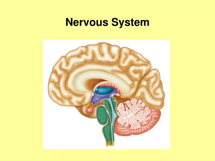
Organization of Nervous System Central Nervous System (CNS) • brain and spinal cord • interprets sensory input and dictates response according to past experience
outside of the CNS Peripheral Nervous System (PNS) • consists of both cranial and spinal nerves • serve as the communication lines that link all parts of the body to the CNS
One of the most important functions of the PNS is to perform reflex actions, which do not involve the brain A reflex is a sudden, rapid, involuntary response to a stimulus
For example: knee jerk reflex (lower leg kicks when tendon is tapped)
withdrawal reflex (hand withdraws if it touches something hot)
pupillary reflex (pupil constricts if a bright light is shown in your eyes)
Knee jerk reflex begins when the patellar tendon is tapped • this generates nerve impulses which travel along sensory neurons to the spinal cord
the spinal cord then transmits impulses through motor neurons to the rectus femoris muscle, which contracts – contraction of the rectus femoris muscle causes extension of the lower leg
The knee jerk reflex involves only 2 types of neurons (sensory and motor), while the withdrawal reflex involves 3 (sensor, association, and motor neurons) • the brain is not involved in the response
information travels over a shorter distance – producing faster reaction times so rapidly in fact that the reflex is completed long before your brain is aware of what happened
you will typically withdraw your hand from something hot before you feel the pain of the burn
Two divisions of Peripheral Nervous System 1. sensory/afferent division • afferent = carrying towards • carries impulses to the CNS from sensory receptors
2. motor division/efferent division • efferent = carrying away • carries impulses from the CNS Two parts: 1. somatic nervous system – voluntary nervous system • regulates skeletal muscles
2. Autonomic Nervous System – involuntary nervous system • includes the cranial and spinal nerves that regulate involuntary actions • regulates smooth/cardiac muscles and glands
Keep in mind that although the ANS functions “automatically”, you can consciously control it for a short period of time Ex. when you hold your breath, you are overriding your ANS – however, you cannot override it indefinitely
The Autonomic Nervous System can be further subdivided into: the Sympathetic Division the Parasympathetic Division
The Sympathetic Division of the ANS regulates involuntary actions during periods of high stress or increased activity For example, the Sympathetic Division: • increases blood pressure, heart rate, breathing rate and blood flow to muscles
dilates the iris of your eyes (increases the amount of light entering the eyes so you can see better) • decreases digestion of food and blood flow to the skin All of these changes prepare your body for activity (“fight or flight”)
The Parasympathetic Division of the Autonomic Nervous System regulates involuntary actions during periods of rest or relaxation For example, the Parasympathetic Division: • decreases blood pressure, heart and breathing rate and blood flow to muscles
constricts the iris of your eyes • increases digestion of food and blood flow to the skin
Most internal organs are regulated by both the sympathetic and parasympathetic divisions in that: • the parasympathetic division dominates when you are relaxed • the sympathetic division dominates when you are under stress
Nervous Tissue • densely packed cells • less than 20% extracellular space
supporting cells (neuroglia) Two types of nerve cells • nerve glue • 6 types (4 in CNS and 2 in PNS)
CNS: astrocytes, microglia, ependymal cells, oligodendrocytes
PNS: satellite cells and schwann cells • various jobs such as chemical production, neuron guidance, neuron growth • increase of action potential conduction Schwann Cell on PNS Motor Neuron satellite cells
nerve cells • Neurons • conduct messages • large, complex cells that have long life-cycles (100+ years) • cells have high metabolic rates
the human brain has an average of 100 billion neurons • amitotic except for olfactory and hippocampal regions
1. cell body Anatomy of Neurons • nuclei/nucleolus surrounded by cytoplasm • also called perikaryon or soma • lacks centrioles • clusters of cell bodies in the CNS are called nuclei • clusters of cell bodies in the • PNS are called ganglia cell body
extend from cell body 2. Processes • in CNS called tracts • in PNS called nerves
Two types of Processes: 1. dendrites – short, tapering, branching extensions • takes messages to the cell body dendrites
2. axons - single process with each neuron • “tail” of neuron, long axon called nerve fiber • generates/transmits nerve impulses axon
white, fatty layer that covers nerve fibers Myelin Sheath • electrically insulates and increases impulse speed
dendrites are always unmyelinated • in PNS formed by Schwann cells • in CNS brain cells that are myelinated are called white matter, unmyelinated cells are gray matter myelin sheath
Structural classification – grouped according to the number of processes extending from the cell body. Classification of neurons 1. multipolar neurons – 3 or more processes • most common (99%) in human CNS
2. bipolar neurons – 2 processes (dendrite and axon) on opposite sides of cell body • rare, found in sense organs such as retina and olfactory mucosa
3. unipolar neurons – single process • more accurately called pseudounipolar neurons • found in ganglia and function as sensory neurons
Functional classification – grouped according to which way the nerve impulse travels to the CNS 1. Sensory (Afferent) neurons – transmit impulses from sensory receptors toward CNS • most are unipolar
2. Motor (Efferent) neurons – transmits impulse away from CNS to effector organs (muscles/glands) • most are multipolar
3. Interneurons (Association) neurons – between motor and sensory neurons • help transmit messages • over 99% of nerves in body • mostly confined to CNS • most are multipolar
highly irritable (responsive to stimuli) Physiology of Neurons • creates response called action potential (nerve impulse) Axon Stimulated Nerve impulse (action potential) Nerve response http://highered.mcgraw-hill.com/sites/0072495855/student_view0/chapter14/animation__the_nerve_impulse.html