Download
1 / 23

E N D
1. DNA Replication • In both prokaryotes and eukaryotes, DNA replication occurs as a prelude to cell division. This DNA replication phase is called the S (synthesis) phase. The two daughter DNA molecules formed from replication eventually become chromosomes in their own right in the daughter cells. • As with all phenomena that involve nucleic acids, the basic machinery of DNA replication depends on complementarity of DNA molecules and on the ability of proteins to form specific interactions with DNA of specific sequences.
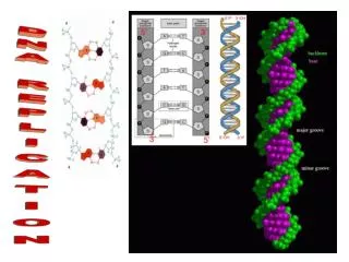
2. The model of Watson and Crick The model of DNA replication proposed by Watson and Crick is based on the hydrogen-bonded specificity of the base pairs. Complementary strands are shown in different colors. The fact that new strands can grow only in the 5’-to-3’ direction adds complexities to the detailed mechanism of replication. If this model is correct, then each daughter molecule should contain one parental nucleotide chain and one newly synthesized nucleotide chain. This prediction has been tested in both prokaryotes and eukaryotes. A little thought shows that there are at least three different ways in which a parental DNA molecule might be related to the daughter molecules. These hypothetical modes are called semiconservative (the Watson-Crick model), conservative, and dispersive
3. Three alternative patterns for DNA replication In semiconservative replication, each daughter duplex contains one parental and one newly synthesized strand. However, in conservative replication, one daughter duplex consists of two newly synthesized strands, and the parent duplex is conserved. Dispersive replication results in daughter duplexes that consist of strands containing only segments of parental DNA and newly synthesized DNA
4. The Meselson-Stahl experiment • In 1958, Matthew Meselson and Franklin Stahl set out to distinguish among the three models. They grew E. coli cells in a medium containing the heavy isotope of nitrogen 15N rather than the normal light (14N) form. This isotope was inserted into the nitrogen bases, which then were incorporated into newly synthesized DNA strands. • After many cell divisions in 15N, the DNA of the cells were well labeled with the heavy isotope. The cells were then removed from the 15N medium and put into a 14N medium; after one and two cell divisions, samples were taken. DNA was extracted from the cells in each of these samples and put into a solution of cesium chloride (CsCl) in an ultracentrifuge.
5. Centrifugation of DNAin a cesium chloride (CsCl) gradient • If cesium chloride is spun in a centrifuge at tremendously high speeds (50,000 rpm) for many hours, the cesium and chloride ions tend to be pushed by centrifugal force toward the bottom of the tube.Ultimately, a gradient of Cs+ and Cl ions is established in the tube, with the highest ion concentration at the bottom. • Molecules of DNA in the solution also are pushed toward the bottom by centrifugal force. But, as they travel down the tube, they encounter the increasing salt concentration, which tends to push them back up owing to the buoyancy of DNA (its tendency to float). Thus, the DNA finally "settles" at some point in the tube where the centrifugal forces just balance the buoyancy of the molecules in the cesium chloride gradient. • The buoyancy of DNA depends on its density (which in turn depends on the ratio of GC to AT base pairs). The presence of the heavier isotope of nitrogen changes the buoyant density of DNA. The DNA extracted from cells grown for several generations on 15N medium can be readily distinguished from the DNA of cells grown on 14N medium by the equilibrium position reached in a cesium chloride gradient. Such samples are commonly called heavy and light DNA, respectively.
6. The proof of the semiconservative model Meselson and Stahl found that, one generation after the heavy cells were moved to 14N medium, the DNA formed a single band of an intermediate density between the densities of the heavy and light controls. After two generations in 14N medium, the DNA formed two bands: one at the intermediate position, the other at the light position. This result would be expected from the semiconservative mode of replication; in fact, the result is compatible with only this mode if the experiment begins with chromosomes composed of individual double helices
7. Harlequin chromosomes • With the use of a more modern staining technique, it is now possible to visualize the semiconservative replication of chromosomes at mitosis. In this procedure, the chromosomes go through two rounds of replication in the presence of bromodeoxyuridine (BUdR), which replaces thymidine in the newly synthesized DNA. The chromosomes are then stained with Giemsa stain, producing the appearance shown. (The light blue lines represent the BUdR-substituted strands.)
8. Visualizing sister chromatids If cells dividing in culture are treated with BrdU during S phase, the cells are fooled into incorporating it — instead of thymidine — into their DNA. One of the properties of the resulting DNA is that it fails to take up stain in a normal way. When cells are allowed to duplicate their chromosomes once in BrdU, the chromosome that appear at the next metaphase stain normally. However, when the cells duplicate their chromosomes a second time in BrdU, one of the sister chromatids that appears at the next metaphase stains normally, while its sister chromatid does not.
9. DNA polymerases • In the late 1950s, Arthur Kornberg successfully identified and purified the first DNA polymerase, an enzyme that catalyzes the replication reaction. • This reaction works only with the triphosphate forms of the nucleotides (such as deoxyadenosine triphosphate, or dATP).
10. DNA polymerases in E. coli • We now know that there are three DNA polymerases in E. coli. The first enzyme that Kornberg purified is called DNA polymerase I or pol I. This enzyme has three activities, which appear to be located in different parts of the molecule: • 1. a polymerase activity, which catalyzes chain growth in the 5’ 3’ direction; • 2. a 3’ 5’exonuclease activity, which removes mismatched bases; and • 3. a 5’ 3’exonuclease activity, which degrades double-stranded DNA. • Subsequently, two additional polymerases, pol II and pol III, were identified in E. coli. Pol II may repair damaged DNA. Pol III, together with pol I, has a role in the replication of E. coli DNA
11. DNA replication fork The complete complex, or holoenzyme, of pol III contains at least 20 different polypeptide subunits, although the catalytic "core" consists of only three subunits. The pol III complex will complete the replication of single-stranded DNA if there is at least a short segment of duplex already present.
12. Prokaryotic origins of replication • E. coli replication begins from a fixed origin, termed oriC, but then proceeds bidirectionally (with moving forks at both ends of the replicating piece). It is 245 bp long and has several components. First, there is a side-by-side, or tandem, set of 13-bp sequences, which are nearly identical. There is also a set of binding sites for a protein, the DnaA protein. An initial step in DNA synthesis is the unwinding of the DNA at the origin in response to binding of the DnaA protein.
13. A replicating E. coli chromosome The DNA has been labeled with 3H-deoxythymidine, and the radioactivity has been detected by overlaying the replicating chromosome with photographic emulsion. The autoradiograph shows that the E. coli chromosome has two replication forks. Although there seem to be two bubbles of replication, actually the point where the two smaller bubbles meet is actually just where two strands of DNA are laying across one another
14. Eukaryotic origins of replication • Bacteria such as E. coli usually require a 40-minute replication-division cycle, but, in eukaryotes, the cycle can vary from 1.4 hours in yeast to 24 hours in cultured animal cells and may last from 100 to 200 hours in some cells. • Eukaryotes have to solve the problem of coordinating the replication of more than one chromosome, as well as replicating the complex structure of the chromosome itself. • In eukaryotes, replication proceeds from multiple points of origin. • Experiments in yeast indicate the existence of about 400 replication origins distributed among the 17 chromosomes, and in humans there are estimated to be more than 10,000 growing forks
15. Replication bubbles in the fruit fly Electron micrograph of replicating DNA in the embryo of the fruit fly D. melanogaster At least 20 different bubbles, therefore with at least 40 different replication forks, can be observed in this electron micrograph (and accompanying drawn representation of the electron micrograph.) The large number of replication origins in eukaryotic chromosomes vs. E. coli's one, enables the slower replication apparatus to copy the larger eukaryotic genome in approximately the same amount of time as the prokaryotic genome is replicated
16. Replication bubbles Electron micrograph of DNA extracted from rapidly dividing nuclei of early D. Melanogaster embryos. The arrows mark replication bubbles; the diameters of DNA chain in both arms of these bubbles indicate that they are double-stranded.
17. Priming DNA synthesis DNA polymerases can extend a chain but cannot start a chain. Therefore, DNA synthesis must first be initiated with a primer, or short oligonucleotide, that generates a segment of duplex DNA. RNA primers are synthesized either by RNA polymerase or by an enzyme termed primase. Primase synthesizes a short (approximately 30 bp long) stretch of RNA complementary to a specific region of the chromosome. The RNA chain is then extended with DNA by DNA polymerase. E. coli primase forms a complex with the template DNA, and additional proteins, such as DnaB, DnaT, Pri A, Pri B, and Pri C. The entire complex is termed a primosome.
18. Leading strand and lagging strand DNA polymerases synthesize new chains only in the 5’ 3’ direction and therefore, because of the antiparallel nature of the DNA molecule, move in a 3’ 5’ direction on the template strand. The consequence of this polarity is that while one new strand, the leading strand, is synthesized continuously, the other, the lagging strand, must be synthesized in short, discontinuous segments. The addition of nucleotides along the template for the lagging strand must proceed toward the template's 5’ end (because replication always moves along the template in a 3’ 5’ directionso that the new strand can grow 5’ 3’). Thus, the new strand must grow in a direction opposite that of the movement of the replication fork.
19. Discontinuous synthesis As fork movement exposes a new section of lagging-strand template, a new lagging-strand fragment is begun and proceeds away from the fork until it is stopped by the preceding fragment. In E. coli, pol III carries out most of the DNA synthesis on both strands, and pol I fills in the gaps left in the lagging strand, which are then sealed by the enzyme DNA ligase. DNA ligases join broken pieces of DNA by catalyzing the formation of a phosphodiester bond between the 5’ phosphate end of a hydrogen-bonded nucleotide and an adjacent 3’ OH group. It is the only enzyme that can seal DNA chains.
20. Steps in DNA synthesis a) The primers for the discontinuous synthesis on the lagging strand are synthesized by primase. b) The primers are extended by DNA polymerase to yield DNA fragments that were first detected by Reiji Okazaki and are termed Okazaki fragments. c) The 5’ 3’exonuclease activity of pol I removes the primers and fills in the gaps with DNA, d) which are sealed by DNA ligase.
22. Other DNA-modifying enzymes • Helicases are enzymes that disrupt the hydrogen bonds that hold the two DNA strands together in a double helix. Among E. coli helicases are the DnaB protein and the Rep protein. The Rep protein may help to unwind the double helix ahead of the polymerase. The unwound DNA is stabilized by the single-stranded binding (SSB) protein, which binds to the single-stranded DNA and retards reformation of the duplex. • The action of helicases during DNA replication generates twists in the circular DNA that need to be removed to allow replication to continue. Circular DNA can be twisted and coiled, much like the extra coils that can be introduced into a rubber band. • This supercoiling can be created or relaxed by enzymes termed topoisomerases. There are two basic types of isomerases. Type I enzymes induce a single-stranded break into the DNA duplex. Type II enzymes cause a break in both strands. In E. coli, topo I and topo III are examples of type I enzymes, whereas gyrase is an example of a type II enzyme.
23. The action of topoisomerases • Untwisting of the DNA strands to open the replication fork causes extra twisting at other regions, and the supercoiling releases the strain of the extra twisting. During replication, gyrase is needed to remove positive supercoils ahead of the replication fork Swivel function of topoisomerase during replication. Extra-twisted (positively supercoiled) regions accumulate ahead of the fork as the parental strands separate for replication. A topoisomerase is required to remove these regions, acting as a swivel to allow extensive replication.