Download
1 / 46
460 likes | 766 Views
Studies with Naphthalimides: An Organic Chemist’s Adventures in the Land of Fluorescence Microscopy. David E. Lewis Department of Chemistry University of Wisconsin-Eau Claire University of Nebraska-Lincoln, March 21, 2005. Who actually did this work?. Lewis Group, 2004

E N D
Studies with Naphthalimides:An Organic Chemist’s Adventures in the Land of Fluorescence Microscopy David E. Lewis Department of Chemistry University of Wisconsin-Eau Claire University of Nebraska-Lincoln, March 21, 2005
Who actually did this work? Lewis Group, 2004 L-R: Glen Gullickson, Grant Sormunen, Jessica Walters, Nick Deprez, Kristy McNitt, DEL Hartsel Group (Biochemistry/Molecular Biology): Scott Hartsel, Lori Scardino, Betsy Ott, Damon Campbell
Fluorescence • Singlet-singlet transitions • Singlet-triplet transition is phosphorescence • Lifetimes typically less than 1 s • Difference between ex and em is known as Stokes shift • Large Stokes shift is desirable to minimize interference from • Scattering • Indigenous fluorescence
Fluorescence spectra of representative 4-amino-1,8-naphthalimides • Large Stokes shifts (≥100 nm) • Large quantum yield of fluorescence • Resistant to photochemical bleaching
Preferred probe properties • high selectivity for the target molecule or organelle. • resistant enough to photochemical degradation under normal illumination conditions to permit the target cell feature to be visualized conveniently. • preferably sufficiently non-toxic to allow live cells to be used for the experiment. • highly fluorescent (i.e. it should have a high quantum yield for fluorescence), so that only small amounts of the dye are needed to visualize the cell target of interest. • large Stokes shift to minimize problems from light scattering by the cell • preferably easy to make from readily available, inexpensive starting materials, and chemically stable to permit long-term storage.
The 4-amino-1,8-naphthalimide fluorophore • Photochemically robust • High quantum yields • Chemically easy to manipulate • Low toxicity • Easily delivered to live cells
Mitochondria are membrane-enclosed organelles distributed through the cytosol of most eukaryotic cells. Their main function is the conversion of the potential energy of food molecules into ATP. Mitochondria have: an outer membrane that encloses the entire structure an inner membrane that encloses a fluid-filled matrix between the two is the intermembrane space the inner membrane is elaborately folded with shelflike cristae projecting into the matrix. a small number (some 5-10) circular molecules of DNA Mitochondria
Key features of the mitochondrion to use in designing a mitochondrial stain • The inner mitochondrial membrane is characterized by • substantial amounts of phosphatidyl serine in the lipid mixture • the presence of a net negative charge on the matrix side of the membrane.
What structural features are needed in the dye? • Delocalized cationic dyes • Sufficient lipohilicity to be membrane-permeant • Cyanines • Mitotracker Green • Triphenylmethane (rhodamine) dyes • reduced dyes • Mitotracker Orange
MitoTracker-type cyanines: 3 resonance contributors with complete octets on all atoms; length of delocalized cation system is 6-7Å rhodamine-type dyes: 4 resonance contributors with complete octets on all atoms; length of delocalized cation system is 9.5Å
A potential new mitochondrial probe n = 6 InstantMito LMT-1 n = 4 InstantMito LMT-2
A potential new mitochondrial probe n = 6 InstantMito LMT-1 n = 4 InstantMito LMT-2 But…
Is a 4-dimethylaminopyridinium ion delocalized enough? • Only 2 resonance contributors with complete octets • Length of conjugated, delocalized cation system is only 4.2Å
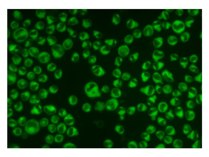
Actually, yes! • Punctate fluorescence – characteristic of mitochondria • Dye is not toxic to cells
Confirming that we are localizing in mitochondria MitoTracker® Red: Commercially available mitochondrion dye Colocalization: Yellow areas show where both dyes occupy the same place in the cell InstantMito LMT-1
Lysosomes: acidic organelles • Lysosomes are roughly spherical bodies bounded by a single membrane. They are manufactured by the Golgi apparatus (pathway 2 in the figure). They contain over 3 dozen different kinds of hydrolytic enzymes including • proteases • lipases • nucleases • polysaccharidases • The pH within the lysosome is about pH 5, substantially less than that of the cytosol (~pH 7.2). All the enzymes in the lysosome work best at an acid pH. This reduces the risk of their digesting their own cell if they should escape from the lysosome.
What structural features are needed in a lysosome probe? • Dyes that have been used for visualizing lysosomes are almost always • weak bases • membrane-permeant in their unprotonated form • tertiary aliphatic amines • Lysotracker Red
A new lysosomal stain InstantLyso LLT-1
Yes! InstantLyso LLT-1 Color epifluorescence image with live THP-1 monocytes at 75 nM and excited with blue light. Colocalization of InstantLyso LLT-1 and Lysotracker Red in live THP-1 cells; yellow represents colocalized probe. 3D reconstruction of a confocal image series using InstantLyso LLT-1
Targeting cholesterol • Plasma membranes are heterogeneous • Membrane partitions into cholesterol-rich and cholesterol-deficient microdomains • The visualization of cholesterol-rich microdomains of plasma membranes (“rafts”) is carried out in a number of ways. • dehydroergosterol • the pentaene antibiotic, filipin • use of labeled cholera toxin subunit B
A new stain for cholesterol-rich microdomains InstantLipo Sep-1 We have also prepared C6 to C18 analogues. These have not all been tested yet, but we know that a minimum of a C8 side chain is required.
Confirming that we are localizing in high-cholesterol domains Vybrant® Alexa Fluor® 594: Current state of the art dye for high cholesterol domains Colocalization: Yellow areas show where both dyes occupy the same place in the cell Instant-Lipo Sep-1 Live THP-1 monocytes
And it works in live foreskin fibroblasts… BODIPY TR C5 ceramide complexed to BSA Colocalization: Yellow areas show where both dyes occupy the same place in the cell Instant-Lipo Sep-1
A putative model for localization cholesterol InstantLipo Sep-1 A 1:1 complex of cholesterol and InstantLipo Sep-1
Golgi apparatus • The Golgi apparatus consists of a stack of membrane-bounded cisternae located between the endoplasmic reticulum and the cell surface. A myriad of enzymes (proteins) are present in the Golgi apparatus to perform its various synthetic activities. So there must be mechanisms • to sort out the processed proteins and send them on to their destinations while • reclaiming processing proteins (e.g., glycosylases) for reuse. • All the details are far from worked out
The accidental discovery: A new stain for Golgi apparatus InstantGolgi McN-1
InstantGolgi McN-1 in fibroblasts BODIPY TR C5 ceramide complexed to BSA Colocalization: Yellow areas show where both dyes occupy the same place in the cell InstantGolgi McN-1 Live foreskin fibroblasts
Lysotracker Red -- the benchmark Lysotracker Red at 75 nM in THP-1 cells. Exposures were taken every 5 seconds (with consistent CCD exposure length) with green excitation cube. Unretouched, unprocessed images. Color is already faded extensively by 7 seconds and is nearly gone by 21 seconds. 7 seconds 21 seconds 35 seconds
InstantLyso LLT-1 InstantLyso LLT-1 at 75 nM in THP-1 cells. Exposures were taken every 30 seconds ( with consistent CCD exposure length 7.5 seconds) with blue excitation cube (490 nm maximum). Each exposure is some increment of 37.5 seconds. We have skipped the middle group of images. Unretouched, unprocessed images. 0 seconds 75 seconds 338 seconds
The comparison… 0 seconds 75 seconds 7 seconds 21 seconds 35 seconds 338 seconds Lysotracker Red InstantLyso LLT-1
InstantLipo Sep-1 InstantLipo Sep-1 at 200 nM in THP-1 cells. Exposures were taken with consistent CCD exposure length with purple excitation cube. Unretouched, unprocessed images. 5 seconds 35 seconds 65 seconds
InstantGolgi McN-1 InstantGolgi McN-1 at 200 nM in THP-1 cells. Exposures were taken with consistent CCD exposure length with purple excitation cube. Unretouched, unprocessed images. 5 seconds 20 seconds 35 seconds
A potentially medium-sensitive probe: fluorescent Tröger’s bases R = n-Bu 57% R = n-C6H13 74% R = n-C8H18 66% Deprez, N.R.; McNitt, K.A.; Petersen, M.E.; Brown, R.G.; Lewis, D.E. Tetrahedron Lett.2005, 46, 2149-2153.
But… It doesn’t cross the cell membrane
Water-soluble neutral probes • Carbohydrate derivatives • Polyether derivatives (e.g. polyethylene glycol derivatives)
Acknowledgments • UW-Eau Claire Office of Research and Sponsored Programs • University of Minnesota NSF-RSEC program