Download
1 / 1
10 likes | 151 Views
STRUCTURAL INSIGHTS INTO FUNCTIONAL MECHANISMS OF NEUROTRANSMITER TRANSPORTERS. II. INFERENCES FROM MOLECULAR MODELS OF ZN-BINDING POCKETS IN hDAT Irache. Visiers* (1) , Lene Norregaard (2), Claus J. Loland (2), Juan A. Ballesteros (1), Ulrik Gether (2), Harel Weinstein (1)

E N D
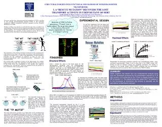
STRUCTURAL INSIGHTS INTO FUNCTIONAL MECHANISMS OF NEUROTRANSMITER TRANSPORTERS. II. INFERENCES FROM MOLECULAR MODELS OF ZN-BINDING POCKETS IN hDAT Irache. Visiers* (1), Lene Norregaard (2), Claus J. Loland (2), Juan A. Ballesteros (1), Ulrik Gether (2), Harel Weinstein (1) (1 )Dpt. Physiology and Biophysics. Mount Sinai School of Medicine, New York. (2) Division of Cellular and Molecular Physiology, Department of Medical Physiology, The Panum Institute, University of Copenhagen, Denmark * e-mail address; irache@inka.mssm.edu An endogenous Zn2+ binding site has been described recently for human dopamine transporter (hDAT)1. This high affinity Zn2+ binding site comprises residues 193His (in the second extracellular loop), 375His (at the top of TM7) and 396Glu (at the top of TM8)2. Zn2+, in micromolar concentrations acts as a potent noncompetitive blocker of dopamine uptake upon binding to this site. An engineered Zn binding site2 indicates that E400C at the top of TM8 is able to coordinate Zn together with 375His, in the absence of 193His, and 396Glu. Based on the structural information contained in Zn binding sites, we build a model of the relative orientation between TM7/TM8 and describe new experiments performed in order to probe the secondary structure of the regions involved. We provide a mechanistic explanation at the molecular level, for the transport inhibition that occurs upon Zn binding. Flexible loop after 375His EXPLORING His 375 to i+5: flexible loop TM8 SEQUENCE ANALYSIS TM8 has two regions with helical periodicity of conservation. The region comprising residues 403 to 405 breaks the helical periodicity of the conservation pattern and presents non-conserved prolines at positions 404 and 405, indicating a disruption in the helical character at this level. 1.25 1 0.75 Conservation 0.5 0.25 V377H, P378H and I379H can substitute H193 in the coordination of Zinc, indicating the flexibility of this region. 0 STRUCTURAL CONSTRAINTS FOR Zn BINDING SITES 387-P 389-L 390-I 392-I 393-I 395-P 398-I 402-P 403-L 417-T 419-G 410-V 391-F 412-F 401-L 413-I 415-L 416-L 418-L 411-F 397-A 399-A 406-A 408-A 414-M 386-G 388-G 400-T 404-S 405-S 409-V 394-Y 396-E 407-W DAT Sequence 402 PPPSSSSPPPPPPPPPPPPPPPPPPPPPPPPPPPPPPP 403 LLLGGGGAAAALLIIIIIIIIVVVVLLLLLLLLFFLLL 404 SSSSSSSSSSSSSSSSSSSSSAAAAPPPSSSSSSSSSS 405 SSSTTTTTTTTPPPPPPPPPPPPPPTTTQQQQQPPPPP Zn(30): 4s2 3d10 Zn2+: 4s0 3d10 FUNTIONAL IMPLICATIONS sp3 hybridization: 4 empty sp3 Covalent bond through the non coupled electron pair in “N”, “O”, or “S”. LIGANDS: His, Glu, Asp, Cys, H2O HISTIDINE: Two possible tautomeric forms: Tautomer : NE2 interacting with metal (70% zinc bound His) Tautomer : His ND1-metal interaction GLU and ASP: COO-groups bind in a monodentate arrangment DISTANCES AND ANGLES: Tightly constrained 3 Thus based on sequence alignment analysis, the predicted secondary structure for TM8 has two non-continuos helical fragments. Although 375His is not likely to reside in a helix, it is possible that Zn binding induces the formation of a helix at the top of TM7, bringing 375His close to 371His. While the segment must be able to adopt such a conformation in the absence of such stabilization, the binding of Zn2+ may reinforce helicity at the external end of TM 7 by stabilizing an otherwise less probable conformation.This induction may entailing a conformational change in the connecting extracellular loop (ECL4), whose implication in transport activity has been reported previously4,5. Sequence alignment analysis reveal important characteristics of this loop: RATIONALE If the transmembrane domain forms an -helix and if this helix includes 375His, it would be expected that a new Zn2+ binding site could be constructed between TM7 and TM8 by removing 193His from the endogenous site and substituting it with a histidine in the i-4 position to 375His. A comprehensive data base search of known structures of naturally occurring Zn2+ binding sites indicates that coordination of Zn2+ between two histidines located in an -helix requires the two histidines to be positioned as i and i-4with i assuming the gauche+ rotamer and i-4 the trans. Coordination of Zn between two His in a helix situated as i and i-3 would require that His adopt a gauche- rotamer which is highly unfavorable. 1. The conservation and hdp pattern of the sequence corresponding to the 4th extracellular loop reveal a short helical fragment from I379 to D385. This segment shows an amphipatic character, where the apolar face is the conserved one suggesting that this short helix has its conserved face in a hydrophobic environment, and its polar not-conserved face toward the aqueous extracellular bath. Graph 1: EXPLORING FROM i-5 TO His 375: helix M371H successfully substitutes H193 in the coordination of Zinc. Any of the other residues from i-5 to His 375 is able to coordinate Zn. 2. A -turn is predicted in the region comprised by residues G386, P387 and G388. These residues are almost 100% conserved which suggests that not only the secondary structure element is important, but the actual residues. Thus the carbonyl backbone groups of G will be highly exposed in a turn so that they may play an important role interacting with other parts of the protein and/or the transported ions. At this point of the project three Zn binding sites were found involving residues in TM7 and TM8: 193His, 375His and 396Glu 375His and 400Cys 371His, 375His and 396Glu If the two coordinating His are localized within a ß-strand they are always separated by only one residue (i and i+2/i-2). TM7 SEQUENCE ANALYSIS Sequence alignment analysis showed that the helix does not continue beyond 375His. Here the models show that residues beyond 375His will be able to coordinate Zn with 396Glu only if those residues are in a flexible loop, which was confirmed experimentally. 1.25 Highly conserved region 3. At least four positions in the 4th extracellular loop are subtype selective, supporting the involvement of this loop in specific extracellular gating mechanisms. 375His is situated close to the extracellular end of TM7, following a highly conserved region with the characteristics of an amphipathic helix. 1 0.75 Conservation 0.5 SUMMARY We have achieved new insight into the structure of a functionally important region located at the external ends of TM 7 and TM 8 of a Na+/Cl--coupled neurotransmitter transporter. In addition to providing new structural information, the insights produced by the ability to engineer several Zn2+ binding sites within this region also defined it as a potential novel site for ligand-mediated, allosteric modulation of transporter function with strong subtype selectivity characteristics. 0.25 0 375-H 381-D 347-I 351-S 352-I 355-L 361-G 368-L 378-P 379-I 385-D 373-Q 353-N 345-D 346-A 371-M 372-A 383-A 349-T 350-T 354-S 356-T 357-S 359-S 360-S 366-S 369-G 376-S 380-G 348-V 363-V 364-V 377-V 382-V 343-Y 344-R 358-F 362-F 365-F 367-F 370-Y 374-K 384-K DAT Sequence 5 Hydrophobic 2.5 0 mean HDP His 375 Hydrophylic -2.5 The tight geometrical constraints for the coordination of Zn, as well as the restriction on the dihedral angles tolerated for two His separated by two residues in an alpha helix, allow us to model the relative orientation of TM7/TM8. Structural criteria and constraints were applied uniformly to be satisfied by all models. A series of cycles of manual refinement produced a final structure compatible with the experimentally derived Zn2+ binding sites, satisfied up to sterically allowed changes in the side chain dihedral angles of the residues involved (but without variation of the relative backbone orientation of the two fragments). -5 375-H 381-D 347-I 351-S 352-I 355-L 361-G 368-L 378-P 379-I 373-Q 353-N 345-D 346-A 371-M 372-A 383-A 385-D 349-T 350-T 354-S 356-T 357-S 359-S 360-S 366-S 369-G 376-S 380-G 348-V 363-V 364-V 377-V 382-V 343-Y 344-R 358-F 362-F 365-F 367-F 370-Y 374-K 384-K DAT Sequence The first three turns of TM7 are predicted to have one face exposed to the lipids, while the last 4 turns are likely to be completely buried in the protein bundle. 375His is the third non conserved polar residue following a strip of 12 conserved residues with a hydrophobicity pattern characteristic of an amphipathic alpha helix. The helix is unlikely to continue after 373Q. 1. Norregaard, L., Frederiksen, D., Nielsen, E. O., and Gether, U. (1998) Embo J 17, 4266-73 2. Loland, C. J., Norregaard, L., and Gether, U. (1999) J Biol Chem 274, 36928-34 3. Alberts, I. L., Nadassy, K., and Wodak, S. J. (1998) Protein Sci 7, 1700-16 4. Penado, K. M., Rudnick, G., and Stephan, M. M. (1998) J Biol Chem 273, 28098-106 5. Smicun, Y., Campbell, S. D., Chen, M. A., Gu, H., and Rudnick, G. (1999) J Biol Chem 274, 36058-64