Download
1 / 22
230 likes | 378 Views
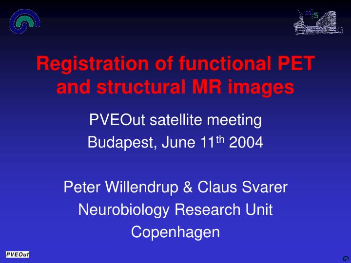
Registration of functional PET and structural MR images. PVEOut satellite meeting Budapest, June 11 th 2004 Peter Willendrup & Claus Svarer Neurobiology Research Unit Copenhagen. Registration needed PVEOut.

E N D
Registration of functional PET and structural MR images PVEOut satellite meeting Budapest, June 11th 2004 Peter Willendrup & Claus Svarer Neurobiology Research Unit Copenhagen
Registration needed PVEOut • Structural MR and functional PET image has to be registered/aligned as the structural information is applied to each voxel in the functional image • As image are coming from same subject only a rigid 6 parameter transformation has to be estimated: • 3 translations (along X, Y and Z axis) • 3 rotations (around X, Y and Z axis) NRU, 2004
What automatic methods are available? • West J, Fitzpatrick JM, Dawant BM, et al. • Headmounted fiducials serves as ``Gold standard'' coregistration between the modalities (MR/CT/PET). • Coregistration parameters are kept for reference, and fiducials are removed from the datasets and replaced by artificial noise. • Methods are tested ``blindly'' - no knowledge of the Gold standard answer. AIR SPM NRU, 2004
Why are the automatic approaches not always a good idea? • These methods are very well suited for registration of images where: • There in the PET image is an equal uptake in all brain regions • There is no inhomogenity variation in the MR images • This is not the case for all receptor PET images, e.g. 5-HT2A altanserin PET images where there are very limited uptake in Cerebellum Limited uptake NRU, 2004
What manual methods have been proposed? Many different approches exist in the litterature • Landmark based: "Graphics applied to medical image registration", G. Q. Maguire, Jr., M. E. Noz, H. Rusinek, et al., Comput Graph Appl, 1991, vol. 11, pp. 20-29. • Surface based: "Accurate three-dimensional registration of CT, PET, and/or MR images of the brain", C. A. Pelizzari, G. T. Y. Chen, D.R. Spelbring, R. R. Wechselbaum, and C-T. Chen, J Comput AssistTomogr, 1989, vol. 13, pp. 20-26. • Image overlay: "Quantitative Comparison of Automatic and Interactive Methods for MRI-SPECT Image Registration of the Brain Based on 3-Dimensional Calculation of Error ”, Pfluger T, Vollmar C et al.: J Nucl Med 2000; 41:1823-1829 • Voxel based: "MRI-PET registration with automated algorithm", R. P. Woods, J. C. Mazziotta, and S. R. Cherry, J Comput Assist Tomogr, 1993, vol. 17, pp. 536-546. NRU, 2004
MARSMultiple Algorithms for Registration of Scans • Modular design • The problem of coregistration can be divided into subtasks • Data selection • Registration • Visualisation / Inspection • Parameter I/O • Reslicing / Re-Interpolation • All subtasks realised by ‘plugins’ - easy inclusion of alternative method • Different registration approaches benefit from shared code NRU, 2004
Main program MARS • This is now included in pvelab NRU, 2004
MARS • Subtask modules • Registration • Interface to Air 5.0 - Roger P. Woods • Interface to SPM 2 - J. Ashburner et. al. • IIO (Interactive Image Overlay) - NRU * • IPS (Interactive Point Selection) - NRU * • Visualisation • Inspect (NRU visualisation program) * • Asterisk-marked will be further explained NRU, 2004
Registration 1: Interactive Image Overlay Translation and rotation of overlay image and surface by keyboard commands NRU, 2004
Inspection of registration Overlay Side by side NRU, 2004
Evaluation study: Setup • Images (5 subjects) • T1 weighted MR images (MPRAGE) • 18F-Altanserin 5HT-2A receptor images • Simulated PET images • Evaluation by 7 volunteers • 3 rounds of MR / Altanserin registration • 1 round of MR / Simulated PET registration • Registration order randomised • Max. one ‘round’ of registrations pr. day • Images also registered using SPM99 and Air 3.0 NRU, 2004
Evaluation study: Simulated PET • Simulated PET datasets • Good: known registration parameters • Bad: “easy” for cost fct. Based methods NRU, 2004
Evaluation study: Altanserin PET • Altanserin PET images • Bad: Lack of gold standard registration method • Good: Real world ‘limited uptake’ images NRU, 2004
Evaluation study • Error measure - Euclidean distance between transformation endpoints • Evaluated for 1% evenly distributed brain voxels. • Mean and std. dev. calculated • Mean transformation realized by 6-parameter estimation to mean of transformed voxels MR PET NRU, 2004
Evaluation study: Simulated PET NRU, 2004
Evaluation study: Altanserin PET NRU, 2004
Evaluation study: Result SPM Air Mean manual No Altanserin binding should be seen in Cerebellum, Rotation problem? Too little binding in Altanserin image, Translation problem? NRU, 2004
Two manual co-registration methods and the interface to two automatic methods have been implemented and incorporated in the PVEOut SW package (pvelab). Four registration methods are included: Interface to SPM 2 (J. Ashburner et. al.) Interface to Air (R. Woods) IIO (NRU) IPS (NRU) For FDG/flow type images, SPM and Air are preferred, with reported errors in the range 2-3 mm. For neuroreceptor type images, with limited binding in areas of the brain, the manual methods can be used and possibly preferred. Measured errors: Registration: Conclusion Simulated images F18-Altanserin images NRU, 2004