Download
1 / 50
500 likes | 603 Views
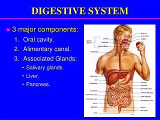
Digestive System. Human Anatomy & Physiology University of Washington PMT. Digestive System Function. Acquires nutrients from environment Anabolism Uses raw materials to synthesize essential compounds Catabolism Decomposes substances to provide energy cells need to function.

E N D
Digestive System Human Anatomy & Physiology University of Washington PMT
Digestive System Function • Acquires nutrients from environment • Anabolism • Uses raw materials to synthesize essential compounds • Catabolism • Decomposes substances to provide energy cells need to function
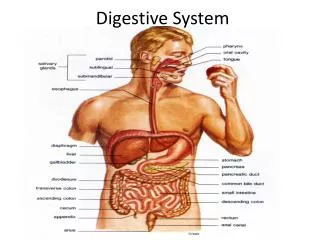
Actions of Digestive (GI) Tract • Ingestion • Occurs when material enters via the mouth • Mechanical Processing • Crushing / Shearing – makes material easier to move through the tract • Digestion • Chemical breakdown of food into small organic compounds for absorption • Secretion • Release of water acids, buffers, enzymes & salts by epithelium of GI tract and glandular organs • Absorption • Movement of organic substrates, electrolytes, vitamins & water across digestive epithelium • Excretion • Removal of waste products from body fluids
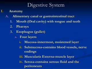
Digestive (GI) Tract • The Digestive Organs and the Peritoneum • Lined with serous membrane consisting of • Superficial mesothelium covering a layer of areolar tissue • Serosa, or visceral peritoneum: • covers organs within peritoneal cavity • Parietal peritoneum: • lines inner surfaces of body wall
Movement of Digestive Materials • By muscular layers of digestive tract • Consist of visceral smooth muscle • Along digestive tract: • Has rhythmic cycles of activities (PERISTALSIS) • Consists of waves of muscular contractions • Move a bolus along the length of the tract • Controlled by pacesetter cells • Surrounding the lumen of the tract • Cells undergo spontaneous depolarization • Triggering wave of contraction through entire muscular sheet
Functions of Oral Cavity • Sensory analysis • Of material before swallowing • Mechanical processing • Through actions of teeth, tongue, and palatal surfaces • Lubrication • Mixing with mucus and salivary gland secretions • Limited digestion • Of carbohydrates and lipids
Esophagus • A hollow muscular tube • About 25 cm (10 in.) long and 2 cm (0.80 in.) wide • Conveys solid food and liquids to the stomach • Begins posterior to cricoid cartilage • Is innervated by fibers from the esophageal plexus
Stomach Function • Major Functions of the Stomach • Storage of ingested food • Mechanical breakdown of ingested food • Disruption of chemical bonds in food material by acid and enzymes • Production of intrinsic factor, a glycoprotein required for absorption of vitamin B12 in small intestine
Digestion in the Stomach • Stomach performs preliminary digestion of proteins by pepsin • Some digestion of carbohydrates (by salivary amylase) • Lipids (by lingual lipase) • Stomach contents • Become more fluid • pH approaches 2.0 • Pepsin activity increases • Protein disassembly begins • Although digestion occurs in the stomach, nutrients are not absorbed there
Small Intestine • 90% of absorption occurs in the small intestine
Small Intestine • The Duodenum • The segment of small intestine closest to stomach • 25 cm (10 in.) long • “Mixing bowl” that receives chyme from stomach and digestive secretions from pancreas and liver • Functions of the duodenum • To receive chyme from stomach • To neutralize acids before they can damage the absorptive surfaces of the small intestine
Small Intestine • The Jejunum • Is the middle segment of small intestine • 2.5 meters (8.2 ft) long • Is the location of most • Chemical digestion • Nutrient absorption • Has few plicae circulares • Small villi
Small Intestine • The Ileum • The final segment of small intestine • 3.5 meters (11.48 ft) long • Ends at the ileocecal valve, a sphincter that controls flow of material from the ileum into the large intestine
Small Intestine • Intestinal Secretions • Watery intestinal juice • 1.8 liters per day enter intestinal lumen • Moisten chyme • Assist in buffering acids • Keep digestive enzymes and products of digestion in solution • Intestinal Movements • Chyme arrives in duodenum • Weak peristaltic contractions move it slowly toward jejunum • Myenteric reflexes • Not under CNS control • Parasympathetic stimulation accelerates local peristalsis and segmentation
Pancreas • Lies posterior to stomach • From duodenum toward spleen • Is bound to posterior wall of abdominal cavity • Is wrapped in thin, connective tissue capsule Functions of the Pancreas • Endocrine cells of the pancreatic islets: • Secrete insulin and glucagon into bloodstream • Exocrine cells: • Acinar cells and epithelial cells of duct system secrete pancreatic juice
Pancreas • Pancreatic Enzymes • Pancreatic alpha-amylase • A carbohydrase • Breaks down starches • Similar to salivary amylase • Pancreatic lipase • Breaks down complex lipids • Releases products (e.g., fatty acids) that are easily absorbed • Pancreatic Enzymes • Nucleases • Break down nucleic acids • Proteolytic enzymes • Break certain proteins apart • Proteases break large protein complexes • Peptidases break small peptides into amino acids • 70% of all pancreatic enzyme production • Secreted as inactive proenzymes • Activated after reaching small intestine
Liver • Hepatocytes • Are liver cells • Adjust circulating levels of nutrients • Through selective absorption and secretion • In a liver lobule form a series of irregular plates arranged like wheel spokes • Many Kupffer cells (stellate reticuloendothelial cells) are located in sinusoidal lining • As blood flows through sinusoids • Hepatocytes absorb solutes from plasma • And secrete materials such as plasma proteins
Liver Function The Physiology of the Liver • Metabolic regulation • Hematological regulation • Bile production
Liver Function • Metabolic Regulation • The liver regulates: • Composition of circulating blood • Nutrient metabolism (carbohydrate, lipid & amino acid) • Waste product removal • Vitamin Storage (A, D, E & K) • Nutrient storage (iron) • Drug inactivation
Liver Function • Composition of Circulating Blood • All blood leaving absorptive surfaces of digestive tract • Enters hepatic portal system • Flows into the liver • Liver cells extract nutrients or toxins from blood • Before they reach systemic circulation through hepatic veins • Liver removes and stores excess nutrients • Corrects nutrient deficiencies by mobilizing stored reserves or performing synthetic activities
Liver Function • Hematological Regulation • Largest blood reservoir in the body • Receives 25% of cardiac output • Functions of Hematological Regulation • Phagocytosis and antigen presentation • Synthesis of plasma proteins • Removal of circulating hormones • Removal of antibodies • Removal or storage of toxins • Synthesis and secretion of bile
Liver Function • The Functions of Bile • Dietary lipids are not water soluble • Mechanical processing in stomach creates large drops containing lipids • Pancreatic lipase is not lipid soluble • Interacts only at surface of lipid droplet • Bile salts break droplets apart (emulsification) • Increases surface area exposed to enzymatic attack • Creates tiny emulsion droplets coated with bile salts
Gallbladder • Is a pear-shaped, muscular sac • Stores and concentrates bile prior to excretion into small intestine • Is located in the fossa on the posterior surface of the liver’s right lobe • The Cystic Duct • Extends from gallbladder • Union with common hepatic duct forms common bile duct
Gallbladder • Functions of the Gallbladder • Stores bile • Releases bile into duodenum, but only under stimulation of hormone cholecystokinin (CCK) • CCK • Hepatopancreatic sphincter remains closed • Bile exiting liver in common hepatic duct cannot flow through common bile duct into duodenum • Bile enters cystic duct and is stored in gallbladder
Coordination of Secretion & Absorption • Intestinal Absorption • It takes about 5 hours for materials to pass from duodenum to end of ileum • Movements of the mucosa increases absorptive effectiveness • Stir and mix intestinal contents • Constantly change environment around epithelial cells
Large Intestine • Is horseshoe shaped • Extends from end of ileum to anus • Lies inferior to stomach and liver • Frames the small intestine • Also called large bowel • Is about 1.5 meters (4.9 ft) long and 7.5 cm (3 in.) wide
Large Intestine Functions • Reabsorption of water • Compaction of intestinal contents into feces • Absorption of important vitamins produced by bacteria • Storage of fecal material prior to defecation
Parts of Large Intestine • The Cecum • Is an expanded pouch • Receives material arriving from the ileum • Stores materials and begins compaction • Appendix • Also called vermiform appendix • Is a slender, hollow appendage about 9 cm (3.6 in.) long • Is dominated by lymphoid nodules (a lymphoid organ)
Parts of Large Intestine • The Colon • Has a larger diameter and thinner wall than small intestine • The wall of the colon • Forms a series of pouches (haustra) • Haustra permit expansion and elongation of colon
Parts of Colon • Ascending Colon • Begins at superior border of cecum • Ascends along right lateral and posterior wall of peritoneal cavity to inferior surface of the liver and bends at right colic flexure (hepatic flexure) • Transverse Colon • Crosses abdomen from right to left; turns at left colic flexure (splenic flexure) • Is supported by transverse mesocolon • Is separated from anterior abdominal wall by greater omentum
Parts of Colon • The Descending Colon • Proceeds inferiorly along left side to the iliac fossa (inner surface of left ilium) • Is retroperitoneal, firmly attached to abdominal wall • The Sigmoid Colon • Is an S-shaped segment, about 15 cm (6 in.) long • Starts at sigmoid flexure • Lies posterior to urinary bladder • Is suspended from sigmoid mesocolon • Empties into rectum
Parts of Large Intestine • The Rectum • Forms last 15 cm (6 in.) of digestive tract • Is an expandable organ for temporary storage of feces • Movement of fecal material into rectum triggers urge to defecate • The anal canal is the last portion of the rectum • Contains small longitudinal folds called anal columns • Anus • Also called anal orifice • Is exit of the anal canal • Has keratinized epidermis like skin
Physiology of the Large Intestine • Absorption in the Large Intestine • Reabsorption of water • Reabsorption of bile salts • In the cecum • Transported in blood to liver • Absorption of vitamins produced by bacteria • Absorption of organic wastes
Physiology of the Large Intestine Three Vitamins Produced in the Large Intestine • Vitamin K (fat soluble): • Required by liver for synthesizing four clotting factors, including prothrombin • Biotin (water soluble): • Important in glucose metabolism • Pantothenic acid: B5 (water soluble): • Required in manufacture of steroid hormones and some neurotransmitters
Physiology of the Large Intestine • Organic Wastes • Bacteria convert bilirubin to urobilinogens and stercobilinogens • Bacteria break down peptides in feces and generate • Ammonia, Indole & skatole, hydrogen sulfide • Bacteria feed on indigestible carbohydrates (complex polysaccharides) • Produce flatus, or intestinal gas, in large intestine
Movements of the Large Intestine • Gastroileal & gastroenteric reflexes • Move materials into cecum while you eat • Movement from cecum to transverse colon is very slow, allowing hours for water absorption • Peristaltic waves move material along length of colon • Segmentation movements (haustral churning) mix contents of adjacent haustra • Movements from transverse colon through rest of large intestine results from powerful peristaltic contractions (mass movements) • Stimulus is distension of stomach and duodenum; relayed over intestinal nerve plexuses • Distension of the rectal wall triggers defecation reflex • Two positive feedback loops • Both loops triggered by stretch receptors in rectum
Digestion • Digestive system handles each nutrient differently • Large organic molecules • Must be digested before absorption can occur • Water, electrolytes, and vitamins • Can be absorbed without processing • May require special transport
Digestion • Digestive Enzymes • Break molecular bonds in large organic molecules • Carbohydrates, proteins, lipids, and nucleic acids • In a process called hydrolysis • Are divided into classes by targets • Carbohydrases break bonds between simple sugars • Proteases break bonds between amino acids • Lipases separate fatty acids from glycerides
Digestion • Water Absorption • Cells cannot actively absorb or secrete water • All movement of water across lining of digestive tract • Involves passive water flow down osmotic gradients