Download
1 / 18
180 likes | 254 Views
Creating Medical Prototypes from MRI data. Jillian Urban Mentor: John T. Tester, Ph.D. Mechanical Engineering. Overview. Project introduction Materials Methods Results Discussion Conclusion. Project Definition. Matlab. Solidworks. Catalyst.

E N D
Creating Medical Prototypes from MRI data Jillian Urban Mentor: John T. Tester, Ph.D. Mechanical Engineering
Overview Project introduction Materials Methods Results Discussion Conclusion
Project Definition Matlab Solidworks Catalyst Reproduce medical prototyping process with the use of basic software tools typically used in an undergraduate mechanical engineering program
Materials • Image files from MRI • Sequenced image files of human anatomy • MRI compiled in AVI format • Dimension SST 768 Prototyping machine • ABS plastic (and support material)
Why incorporate the two? MRI compiled to AVI original MRI format is proprietary AVI universal format portable for medical personnel Create solid model of anatomy
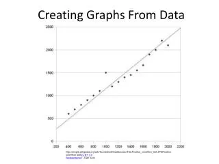
Point cloud to STL • Arrange pixels in x-, y-, and z- coordinates • Stack the images for 3D • Point cloud to mesh using Scanto3D • STL file created for Rapid Prototyping 12
Results Meshed skull outer surface 13
Results Meshed jaw with outer and inner surface 14
Discussion Pixel Detection Advantages Outer layer Small features Disadvantages Not sufficient for production of inner and outer layers combined I.E., SHELLS Canny Advantages Noise reduction Production of shell structures in CAD Accurate cavity bone location Disadvantages More smoothing for precise meshing 15
Discussion Scanto3D Advantages Mesh creation from point cloud STL creation Disadvantages Inaccurate meshing of interior complex bone cavities Less control of surface creation 16
Conclusion Three-step method of processing AVI files Most complex bone structure: the skull Lessons learned Scanto3D meshing sufficient for external surface meshing of a solid form Small medical practices can produce solid models of simple bone structures using conventional computer engineering tools Need more work for complex bone shell-type structures 17
Questions Special thanks to NAU NASA Space Grant Internship Program, Flagstaff Medical Center, and Dr. John Tester 18