Download

1 / 24
240 likes | 393 Views
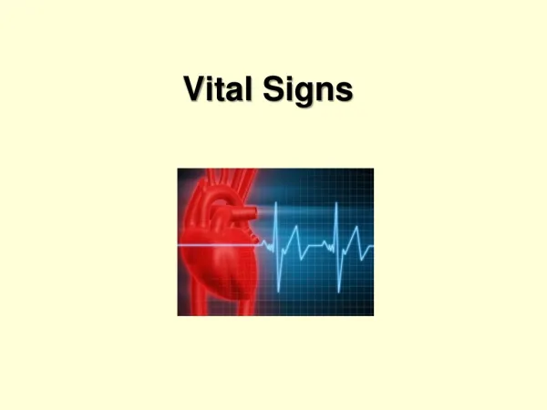
VITAL SIGNS. BY Georges Metellus, M.D., M.P.H. VITAL SIGNS. Are considered the baseline indicators of a patient’s health status. They may be measured early in the physical examination. Pulse Respiration Blood Pressure Temperature. Pulse.

E N D
VITAL SIGNS BY Georges Metellus, M.D., M.P.H
VITAL SIGNS Are considered the baseline indicators of a patient’s health status. They may be measured early in the physical examination. • Pulse • Respiration • Blood Pressure • Temperature
Pulse • The pulse may provide information about the rate, strength, and rhythmicity of the heartbeat. • The pulse may be palpated in several different areas. The nine major “pulse points” are named after the arteries over which they are felt. • To feel a pulse, you must place the pads of your second and third fingers over an artery that lies near the surface of the body and over a bone or a firm base.
Pulse Points • 1. Over the superficial temporal artery in front of the ear. • 2. The common carotid artery in the neck along the front edge of the sternocleidomastoid muscle. • 3. Over the facial artery at the lower margin of the mandible at a point below the corner of the mouth. • 4. In the axilla over the axillary artery • 5. Over the brachial artery at the bend of the elbow along the inner or medial margin of the biceps brachii muscle • 6. At the radial artery at the wrist (radial pulse). It is the most frequently monitored and easoly accessible in the body • 7. Over the femoral artery in the groin. • 8. At the popliteal artery behind and just proximal to the knee • 9. At the dorsalispedis artery on the front surface of the foot, just below the bend of the ankle joint
Arterial pulse abnormalities Type/description Associated conditions • Pulsusalternans: regular rate; amplitude varies from beat to beat with weak and strong beats • Bigeminal pulse: Two beats in rapid succession followed by longer interval; easily confused with alternating pulse. • Pulsusbisferiens: Two strong systolic peaks separated by a midsystolic dip. • Left ventricular failure • Regularly occuring ventricular premature beats • Aortic regurgitation alone or with stenosis
Arterial Pulse Abnormality cont’ Type/description Associated conditionS • Bounding pulse: increase pulse pressure; contour may have rapid rise, brief peak, rapid fall. • Bradycardia: rate <60 • Artherosclerosis, aortic rigidity, patent ductucarteriosus, fever, anemia, hyperthyroidism, anxiety, exercise • Hypothermia, Hypothyroidism, drug intoxication, impaired cardiac conduction, exellent physical conditioning
Arterial pulse Abnormalities cont’ Type/Description Associated conditions • Paradoxic pulse: Amplitude decreases on inspiration • Pulsusdifferens: Unequal pulses between left and right extremities • Tachycardia: Rate over 100 • Chronic obstructive disease, constrictive pericarditis, peircardial effusion • Impaired circulation, usually from unilateral local obstruction • Fever, hyperthydoidism, anemia, shock, Heart disease, anxiety, exercise
Arterial pulse abnormalities cont’ Type/description Associated conditions • Trigeminal pulse: three beats followed by a pause. • Water-hammer pulse (corrigan pulse): Jerky pulse with full expansion followed by sudden collapse • Often benign, such as after exercise; but may occur withcardiomyopathy, severe ventricular hypertrophy, severe aortic stenosis, dysfunctional right ventricle. • Aortic regurgitation
Blood pressure • Blood pressure is the pressure or “push” of blood as it flows through the circulatory system. It is a peripheral measurement of cardiovascular function. • Indirect measures of blood pressure are made with a stethoscope and a sphygmomanometer (aneroid or mercury) or with electronic sphygmomanometers which do not require the use of a stethoscope
Factors That Influence Blood Pressure • Blood Volume • The strength of each heart contraction • Heart rate • The thickness of blood (viscosity) • Rigidity of the arteries
Blood Pressure and blood volume • The larger the volume of blood in the arteries, the more pressure the blood exerts on the walls of the arteries. • The less blood in the arteries, the lower the blood pressure tends to be. (Hemorrhage demonstrates this relation between blood volume and blood pressure)
Strength of Heart Contraction and Blood Pressure • A stronger heartbeat increases blood pressure and a weaker beat decreases it. • Cardiac output is also influenced by the strength of the contraction of the heart
Heart Rate and Blood Pressure • The rate of the heart also affect arterial Blood pressure. When the heart beats faster, more blood enters the aorta, therefore the arterial blood volume and blood pressure would increase. • The stroke volume is to be considered because it might determine whether or not the blood pressure is going to change in one way or another
Blood Viscosity and Blood pressure • If blood becomes less viscous than normal, blood pressure decreases. (if a person suffers a hemorrhage, fluid moves into the blood from the interstitial fluid. This dilutes the blood and decreases its viscosity, and blood pressure) • In a condition called Polycythemia, the number of red blood cells increases beyond normal and thus increases blood viscosity. This in turn increases blood pressure.
Increase of Arteriolar Resistance and Blood Pressure Increased arteriolar resistance is the most common cause of hypertension. This increase may occur secondarily to: 1. Endocrine causes: Tumor of the adrenal medulla ( Pheochromocytomas) produces epinephrine and norepinerphrine and may give rise to paroxysmal form of hypertension 2. Renal Causes: a) Renal parenchyma: Chronic glomerulonephritis Pielonenephritis Polycystic disease B) Renal vasculature vascular lesions due to congenital or acquired malformation of the renal artery or to small vessels disease as such in lupus erythematosus. 3. Essential Hypertension: is the most cause of a pathologically elevated blood pressure. The disease shows a marked familial tendency, and it appears commonly in middle-aged people. It is one of the most common causes of left ventricular Hypertrophy.
Hypotension Hypotension results from: 1. Decrease of cardiac output: In Addison’s disease, myocarditis, myocardial infarction, pericarditis with effusion, and following Hemorrhage 2. Decrease in peripheral resistance Vasomotor collapse, may occur in: Pneumonia Septicemia Acute Adrenal insufficiency (waterhouse-Frederichsen syndrome) Drug intoxication ( a sudden drop in blood pressure should be regarded as a grave sign)
Respiration • Respiration means exchange of gases( oxygen and carbon dioxide) between a living organism and its environment • Respirations are counted and evaluated by inspection. Observe the rise and the fall of the patient’s chest and the ease with which breathing is accomplished. Count the number of respiratory cycles (inspiration and expiration) per minute. • Observe the regularity and rhythm of the breathing pattern. • Note the depth of respirations and whether the patient uses accessory muscles.
Respiratory Patterns • Tachypnea:Is a persistent respiratory rate approaching 25 respirations per minute. Certain patients with fibrosis of the lung, pulmonary edema, pleural disease, or rib cage fixation may breathe rapidly and shallowly. Other patients may increase the minute ventilation to accommodate an increased gas exchange that is necessitated by exercise,fever,hypermetabolic states, or anxiety
Respiratory Patterns: Cont’ • Bradypnea:rate slower than 12 respiration per minute. May indicate neurologic ( i.eintracraneal pressure: hemorrhage, tumor) or electrolyte disturbance, infection or a sensible response to protect against the pain of pleurisy or other irritative phenomena (it may also mean splendid level of cardiorespiratory fitness) • Kussmaul Respiration: deep, rapid and labored respiration associated with metabolic acidosis. May indicate decompensated diabetes with profund acidosis; renal diseases or drug causing acidosis. Diseases of the central nervous system, such as meningitis, may increase minute ventilation
Respiratory Patterns: cont’ • Cheyne-Stokes respiration: A regular periodic pattern of breathing, with intervals of apnea followed by a crescendo/decrescendo sequence of respiration. It occurs in patients who are seriously ill, particularly those with brain damage at the cerebral level or with drug-caused respiratory compromise. • Biot Respiration: Consists of somewhat irregular respirations varying in depth and interrupted by intervals of apnea. It is usually associated with severe and persistent increase intracranial pressure, respiratory compromise resulting from drug poisoning, or brain damage at the level of the medulla.
Temperature • The assessment of body temperature may often provide an important clue to the severity of a patient’s illness. Temperature measurement can be accomplished through several different routes, most commonly: oral, rectal, axillary, tympanic membrane (less common) In the case of bacterial infection it may well be the most critical diagnostic indicator, especially with infants, toddlers, and the elderly
Temperature Cont’ Important conditions related to body temperature: 1. Fever: Is an unsually high body temperature associated with a systemic inflamatory response. In the case of infections, chemical called Pyrogens cause the thermostatic control centers of the hypothalamus to produce fever. 2. Malignant hyperthermia: Is an inherited condition characterized by an abnormally increased body temperature and muscle rigidity when exposed to certain anesthestics 4. Heat exhaustion: Occurs when the body loses a large amount of fluid resulting from heat-loss mechanism. This usually happens when environmental temperatures are high. The loss of water and electrolytes can cause weakness, vertigo, nausea, heat cramps and possibly loss of consciousness
Important conditions related to body temperature • Heatstroke or sunstroke: is a severe condition resulting from the inability of the body to maintain a normal temperature in an extremely warm environment. Such thermoregulatory failure may result from factors such as old age, disease, drugs that impair thermoregulation, or simply overwhelming elevated envinonmental temperatures.
Temperature Cont’ • Hypothermia:is the inability to maintain a normal body temperature in extremely cold environments. Hypothermia is characterized by body temperature lower than 35C (95F), shallow and slow respirations, and a faint, slow pulse. • Frostbite: is local damage to tissues caused by extremely low temperatures. Necrosis and even gangrene can result from frostbite.