Download
1 / 1
10 likes | 109 Views
Characterizing the microbial diversity of hydrocarbon contaminated soil via capillary and pyrosequencing By KC VerHage SUNY Oswego Department Of Biological Sciences and Uni. Of Calcutta; B.C. Guha Centre for Biotechnology. RESULTS AND DISCUSSION. ABSTRACT.

E N D
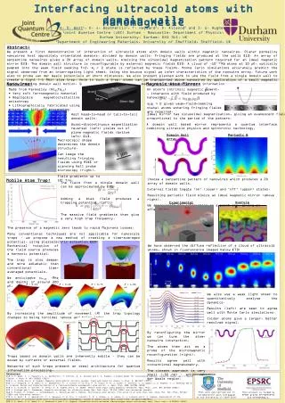
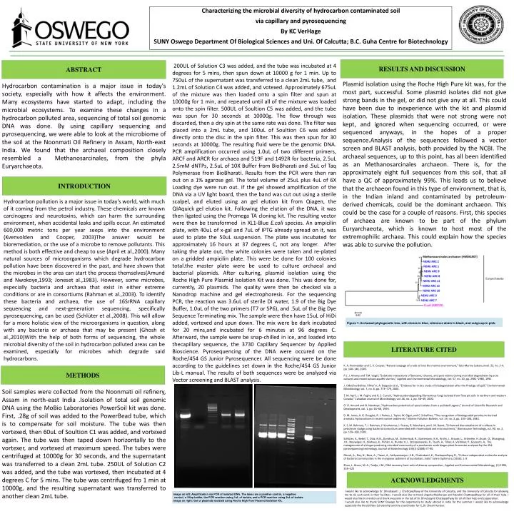
Characterizing the microbial diversity of hydrocarbon contaminated soil via capillary and pyrosequencing By KC VerHage SUNY Oswego Department Of Biological Sciences and Uni. Of Calcutta; B.C. Guha Centre for Biotechnology RESULTS AND DISCUSSION ABSTRACT 200UL of Solution C3 was added, and the tube was incubated at 4 degrees for 5 mins, then spun down at 10000 g for 1 min. Up to 750uL of the supernatant was transferred to a clean 2mL tube, and 1.2mL of Solution C4 was added, and votexed. Approximately 675uL of the mixture was then loaded onto a spin filter and spun at 10000g for 1 min, and repeated until all of the mixture was loaded onto the spin filter. 500UL of Soultion C5 was added, and the tube was spun for 30 seconds at 10000g. The flow through was discarded, then a dry spin at the same rate was done. The filter was placed into a 2mL tube, and 100uL of Soultion C6 was added directly onto the disc in the spin filter. This was then spun for 30 seconds at 10000g. The resulting fluid were be the genomic DNA. PCR amplification occurred using 1.0uL of two different primers, ARCF and ARCR for archaea and 519F and 1492R for bacteria, 2.5uL 2.5mM dNTPs, 2.5uL of 10X Buffer from BioBharati and .5uL of Taq Polymerase from BioBharati. Results from the PCR were then ran out on a 1% agarose gel. The total volume of 25uL plus 4uL of 6X Loading dye were run out. If the gel showed amplification of the DNA via a UV light board, then the band was cut out using a sterile scalpel, and eluted using an gel elution kit from Qiagen, the QIAquick gel elution kit. Following the elution of the DNA, it was then ligated using the Promega TA cloning kit. The resulting vector were then be transformed in XL1-Blue E.coli species. An ampicilin plate, with 40uL of x-gal and 7uL of IPTG already spread on it, was used to plate the 50uL suspension. The plate was incubated for approximately 16 hours at 37 degrees C, not any longer. After taking the plate out, the white colonies were taken and re-plated on a gridded ampicilin plate. This were be done for 100 colonies total.the master plate were be used to culture archaeal and bacterial plasmids. After culturing, plasmid isolation using the Roche High Pure Plasmid Isolation Kit was done. This was done for, currently, 20 plasmids. The quality were then be checked via a Nanodrop machine and gel electrophoresis. For the sequencing PCR, the reaction was 3.6uL of sterile DI water, 1.9 of the Big Dye Buffer, 1.0uL of the two primers (T7 or SP6), and .5uL of the Big Dye Sequence Terminating mix. The sample were then have 15uL of HiDi added, vortexed and spun down. The mix were be dark incubated for 20 mins,and incubated for 6 minutes at 96 degrees C. Afterward, the sample were be snap-chilled in ice, and loaded into thecapillary sequence, the 3730 Capillary Sequencer by Applied Bioscience. Pyrosequencing of the DNA were occured on the Roche/454 GS Junior Pyrosequencer. All sequencing were be done according to the guidelines set down in the Roche/454 GS Junior Lib-L manual. The results of both sequences were be analyzed via Vector screening and BLAST analysis. Plasmid isolation using the Roche High Pure kit was, for the most part, successful. Some plasmid isolates did not give strong bands in the gel, or did not give any at all. This could have been due to inexperience with the kit and plasmid isolation. These plasmids that were not strong were not kept, and ignored when sequencing occurred, or were sequenced anyways, in the hopes of a proper sequence.Analysis of the sequences followed a vector screen and BLAST analysis, both provided by the NCBI. The archaeal sequences, up to this point, has all been identified as an Methanosarcinales archaeon. There is, for the approximately eight full sequences from this soil, that all have a QC of approximately 99%. This leads us to believe that the archaeon found in this type of environment, that is, in the Indian inland and contaminated by petroleum-derived chemicals, could be the dominant archaeon. This could be the case for a couple of reasons. First, this species of archaea are known to be part of the phylum Euryarchaeota, which is known to host most of the extremophilic archaea. This could explain how the species was able to survive the pollution. Hydrocarbon contamination is a major issue in today's society, especially with how it affects the environment. Many ecosystems have started to adapt, including the microbial ecosystems. To examine these changes in a hydrocarbon polluted area, sequencing of total soil genomic DNA was done. By using capillary sequencing and pyrosequencing, we were able to look at the microbiome of the soil at the Noonmati Oil Refinery in Assam, North-east India. We found that the archaeal composition closely resembled a Methanosarcinales, from the phyla Euryarchaeota. INTRODUCTION Hydrocarbon pollution is a major issue in today's world, with much of it coming from the petrol industry. These chemicals are known carcinogens and neurotoxins, which can harm the surrounding environment, when accidental leaks and spills occur. An estimated 600,000 metric tons per year seeps into the environment (Kvenvolden and Cooper, 2003)The answer would be bioremediation, or the use of a microbe to remove pollutants. This method is both effective and cheap to use (April et al.,2000). Many natural sources of microorganisms which degrade hydrocarbon pollution have been discovered in the past, and have shown that the microbes in the area can start the process themselves(Amund and Nwokoye,1993; Joneset al.,1983). However, some microbes, especially bacteria and archaea that exist in either extreme conditions or are in consortiums (Rahman et al.,2003). To identify these bacteria and archaea, the use of 16SrRNA capillary sequencing and next-generation sequencing, specifically pyrosequencing, can be used (Schlüter et al.,2008). This will allow for a more holistic view of the microorganisms in question, along with any bacteria or archaea that may be present (Ghosh et al.,2010)With the help of both forms of sequencing, the whole microbial diversity of the soil in hydrocarbon polluted areas can be examined, especially for microbes which degrade said hydrocarbons. Figure 1: Archaeael phylogenetic tree, with clones in blue, reference strain in black, and outgroup in pink] LITERATURE CITED K. A. Kvenvolden and C. K. Cooper, “Natural seepage of crude oil into the marine environment,” Geo-Marine Letters,mvol. 23, no. 3-4, pp. 140–146, 2003. P. J. J. Alvarez and T.M. Vogel, “Substrate interactions of benzene, toluene, and para-xylene during microbial degradation by pure cultures and mixed culture aquifer slurries,” Applied and Environmental Microbiology, vol. 57, no. 10, pp. 2981–2985, 1991. J. I.Medina-Bellver, P.Mar´ın, A. Delgado et al., “Evidence for in situ crude oil biodegradation after the Prestige oil spill,” Environmental Microbiology, vol. 7, no. 6, pp. 773–779, 2005. T. M. April, J. M. Foght, and R. S. Currah, “Hydrocarbondegrading filamentous fungi isolated from flare pit soils in northern and western Canada,” Canadian Journal of Microbiology, vol. 46, no. 1, pp. 38–49, 2000. O. O. Amund and N. Nwokoye, “Hydrocarbon potentials of yeast isolates from a polluted Lagoon,” Journal of Scientific Research and Development, vol. 1, pp. 65–68, 1993. D. M. Jones, A. G. Douglas, R. J. Parkes, J. Taylor, W. Giger, and C. Schaffner, “The recognition of biodegraded petroleum-derived aromatic hydrocarbons in recent marine sediments,” Marine Pollution Bulletin, vol. 14, no. 3, pp. 103–108, 1983. K. S. M. Rahman, T. J. Rahman, Y. Kourkoutas, I. Petsas, R. Marchant, and I. M. Banat, “Enhanced bioremediation of n-alkane in petroleum sludge using bacterial consortium amended with rhamnolipid and micronutrients,” Bioresource Technology, vol. 90, no. 2, pp. 159–168, 2003. Schlüter, A., Bekel, T., Diaz, N.N., Dondrup, M., Eichenlaub, R., Gartemann, K.H., Krahn, I., Krause, L., Krömeke, H.,Kruse, O., Mussgnug, J.H., Neuweger, H., Niehaus, K., Pühler, A., Runter, K.J., Szczepanowski, R., Tauch, A., Tilker, A.,Viehöver, P., Goesann, A,. The metagenome of a biogas-producing microbial community of a production-scale biogas plant fermenter analysed by the 454-pyrosequencing technology. Journal of Biotechnology 136(1) (2008):77-90. Ghosh, A., Dey, N., Bera, A., Tiwari, A., Sathyaniranjan, K.B., Chakrabarti, K., Chattopadhyay, D., “Culture independent molecular analysis of bacterial communities in the mangrove sediment of Sundarban, India” Saline Systems 6, (2010): 1-9 Zhou, J., Bruns, M. A., Tiedje, J.M., DNA recovery from soils of diverse composition., Applied and Environmental Microbiology, (2) 1996, 316–322 METHODS Soil samples were collected from the Noonmati oil refinery, Assam in north-east India .Isolation of total soil genomic DNA using the MoBio Laboratories PowerSoil kit was done. First, .28g of soil was added to the PowerBead tube, which is to compensate for soil moisture. The tube was then vortexed, then 60uL of Soultion C1 was added, and vortexed again. The tube was then taped down horizontally to the vortexer, and vortexed at maximum speed. The tubes were centrifuged at 10000g for 30 seconds, and the supernatant was transferred to a clean 2mL tube. 250UL of Solution C2 was added, and the tube was vortexed, then incubated at 4 degrees C for 5 mins. The tube was centrifuged fro 1 min at 10000g, and the resulting supernatant was transferred to another clean 2mL tube. ACKNOWLEDGMENTS I would like to acknowledge Dr. Dhrubajyoti .J. Chattopadhyay of the University of Calcutta, and the University of Calcutta for allowing me to do such work in their facilities. I would also like to thank Argyha Mukherjee and Nandini Chattopadhyay for all of their help. I would also like to mention and thank everyone in the lab of Dr. Dhrubajyoti Chattopadhyay for all of their help and cooperation. I would also like to thank SUNY Oswego for the opportunity to study abroad in India for the summer. I would like to acknowledge especially the Possibilities Scholarship and the coordinator for it, Dr. Shashi Kanbur. Image on left: Amplification via PCR of isolated DNA. The lanes are a positive control, a negative control, a 1Kbp ladder, the PCR reaction using 1uL of isolate, and a PCR reaction using 2uL of isolate Image on right: Gel of plasmids isolated using Roche High Pure Plasmid Isolation Kit.