Download
1 / 84
840 likes | 853 Views
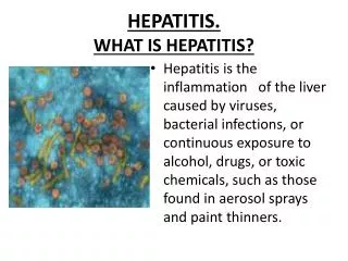
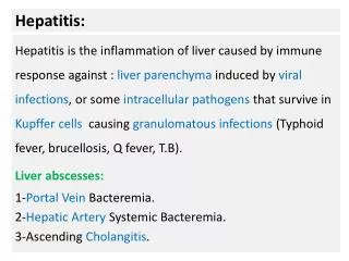
HEPATITIS. HEPATITIS = inflammation of liver. HEPATITIS - causes. ACUTE: Viral hepatitis Non - viral infection Alcohol Toxins Drugs Ischemic hepatits Autoimmune Metabolic diseases. CHRONIC: Viral hepatitis Alcohol Drugs Non-alcoholic steatohepatitis Autoimmune Heredity.

E N D
HEPATITIS- causes ACUTE: • Viral hepatitis • Non-viral infection • Alcohol • Toxins • Drugs • Ischemic hepatits • Autoimmune • Metabolic diseases CHRONIC: • Viral hepatitis • Alcohol • Drugs • Non-alcoholic steatohepatitis • Autoimmune • Heredity
HEPATITIS-symptoms ACUTE: • Malaise • Muscle and join ache • Fever • Nausea or vomiting • Loss of apetite • Abdominal pain • Dark urine • Jaundice CHRONIC: • Malaise, tiredness, weakness • Weight loss • Peripheral oedema • Ascites
VIRAL HEPATITIS • The term viral hepatitis is used to describe infection of the liver caused by hepatotropic viruses. • Currently there are 5 main varieties of these viruses
ETIOLOGIC CLASSIFICATION • Based on the etiologic agent, viral hepatitis is currently classified into 6 etiologic types—hepatitis A, hepatitis B, hepatitis C, hepatitis D, hepatitis E and hepatitis G.
Hepatitis A • Infection with HAV causes hepatitis A (infectious hepatitis). • Hepatitis A is usually a benign, self-limiting disease and has an incubation period of 15-45 days. • Spread:by faeco-oral route & is related to close personal contact such as in overcrowding, poor hygiene and poor sanitation. • Most frequently affected age is 5-14 years; adults are often infected by spread from children.
HEPATITIS A VIRUS (HAV) • Small, 27 nm diameter, icosahedral non-enveloped, single-stranded RNA virus. • Virus is present in the liver cells, bile, stool and blood during the incubation period and in pre-icteric phase but viral shedding diminishes after the onset of jaundice. • Chroniccarriers have not been identified for HAV infection.
PATHOGENESIS • Poorly understood. • An immunologic basis is suspected.
Markers are: 1. IgM anti-HAV antibodyappears in the serum at the onset of symptoms of acute hepatitis A. 2. IgG anti-HAV antibody is detected in the serum after acute illness and remains detectable indefinitely. It gives life-long protective immunity against reinfection with HAV.
Hepatitis B • Hepatitis B (serum hepatitis) caused by HBV infection. • Longer incubation period (30-180 days) • Occur at any age.
Transmitted parenterally such as in • recipients of blood and blood products • intravenous drug addicts • patients treated by renal dialysis • hospital workers exposed to blood • by intimate physical contact such as from mother to child • sexual contact
HBV infection causes more severe form of illness that includes: acute hepatitis B, chronic hepatitis, progression to cirrhosis, fulminant hepatitis and an asymptomatic carrier stage. • HBV plays some role in the development of hepatocellular carcinoma.
HEPATITIS B VIRUS (HBV) • DNA virus • Electron microscopic studies on serum of patients infected with HBV show 3 forms of viral particles of 2 sizes: small (spheres and tubules/filaments) and large (spheres). • Genomic structure of HBV is quite compact and complex. • HBV DNA consists of 4 overlapping genes which encode for multiple proteins.
PATHOGENESIS • Several immunological markers (serologic as well as viral), and molecular and morphologic evidence suggest that hepatocyticdamage is initiated by virus-infected CD8+T cytotoxic cells.
Serologic and viral markers Detected in sera and hepatocytes of infected individual 1. HBsAg. • Australia antigen • HBsAg appears early in the blood after about 6 weeks of infection and its detection is an indicator of active HBV infection. • It usually disappears in 3-6 months. • Its persistence for more than 6 months implies a carrier state. • HBsAg may also be demonstrated in the cell membrane of hepatocytes of carriers and chronic hepatitis patients by Orceinstaining (orange positivity) but not in the hepatocytes during acute stage of illness.
2. Anti-HBs. • Specific antibody to HBsAg in serum • Appears late, about 3 months after the onset. • Anti-HBs response may be both IgM and IgG type. • Itpersists for life providing protection against reinfection with HBV. 3. HBeAg. • HBeAgderived from core protein is present transiently (3-6 weeks) during an acute attack. • Its persistence beyond 10 weeks is indicative of development of chronic liver disease and carrier state.
4. Anti-HBe. • Antibody to HBeAg called anti-HBeappears after disappearance of HBeAg. • Seroconversionfrom HBeAgto anti-HBe during acute stage of illness is a prognostic sign for resolution of infection. 5. HBcAg. • HBcAgderived from core protein cannot be detected in the blood. • HBcAgcan be demonstrated in the nuclei of hepatocytes in carrier state and in chronic hepatitis patients by Orcein staining but not in the liver cells during acute stage.
6. Anti-HBc. • Detected in the serum of acute hepatitis B patients during pre-icteric stage. • IgManti-HBc persists for 4-6 months and is followed later by IgG anti-HBc. • Detection of high titre of IgM anti-HBc is indicative of recent acute HBV infection • Elevated level of IgG anti-HBc suggests HBV infection in the remote past. 7. HBV-DNA. • Detection of HBV-DNA by molecular hybridisation using the Southern blot technique is the most sensitive index of hepatitis B infection. • It is present in presymptomaticphase and transiently during early acute stage.
Hepatitis D • Infection with delta virus (HDV) in the hepatocyte nuclei of HBsAg-positive patients is termed hepatitis D. • HDV is a defective virus for which HBV is the helper. • Hepatitis D develops when there is concomitant hepatitis B infection. • HDV infection and hepatitis B may be simultaneous (coinfection), or HDV may infect a chronic HBsAgcarrier (superinfection).
High-risk individuals for HDV infection are the same as for HBV infection i.e. • intravenous drug abusers • Homosexuals • transfusion recipients • health care workers
HEPATITIS DELTA VIRUS (HDV) • Small single-stranded RNA particle with a diameter of 36 nm. • It is double-shelled—the outer shell consists of HBsAg and the inner shell consists of delta antigen provided by a circular RNA strand. • It is highly infectious and can induce hepatitis in any HBsAg-positive host.
Markers for HDV infection include: 1. HDV identification in the blood and in the liver cell nuclei. 2. HDAgdetectable in the blood and on fixed liver tissue specimens. 3. Anti-HD antibody in acute hepatitis which is initially IgM type and later replaced by IgG type anti-HD antibody which persists for life to confer immunity against reinfection.
Hepatitis C • Diagnosis was earlier made after exclusion of infection with other known hepatitis viruses and was initially designated non-A, non-B (NANB) hepatitis. • Now called hepatitis C.
Hepatitis C infection is acquired by blood transfusions, blood products, haemodialysis, parenteral drug abuse and accidental cuts and needle-pricks in health workers. • Incubation period of 20-90 days (mean 50 days). • Persistence of infection and chronic hepatitis are the key features of HCV. • Late consequences occurrence of cirrhosis after 5 to 10 years and progression to hepatocellular carcinoma • HCV is considered more important cause of chronic liver disease worldwide than HBV.
HEPATITIS C VIRUS (HCV) • HCV is a single-stranded, enveloped RNA virus, having a diameter of 30-60 nm. HCV genome has about 3000 amino acids. • The genomic organisation of HCV shows a 5’ terminal end, C (capsid) region and the envelope regions E1 and E2 in the exons.
Viral proteins result in corresponding serologic and virologic markers for HCV infection 1. Anti-HCV antibodies. Three generations of anti-HCV IgGassays are available: i) First generation antibodies are against C100-3 region proteins and appear 1 to 3 months after infection. ii) Second generation antibodies are against C200 and C33c proteins and appear about one month earlier than the first generation. iii) Third generation antibodies are against C22-3 and NS-5 region proteins and are detected even earlier. 2. HCV-RNA. • PCR technique which can be detected within a few days after exposure to HCV infection, much before appearance of anti-HCV • Persists for the duration of HCV infection.
PATHOGENESIS • Cell-mediated immune mechanism and production of antiviral cytokines by T-lymphocytes certainly play a role in hepatocytic injury due to HCV. • Crossreactivitybetween viral antigens and host autoantibodies to liver-kidney microsomal antigen (anti- LKM) have been reported in a subset of patients that explains the association of autoimmune hepatitis and HCV hepatitis.
Hepatitis E • Enterically-transmitted virus, previously labelled as epidemic or enterically transmitted variant of non- A non-B hepatitis. • Infection is generally acquired by contamination of water supplies such as after monsoon flooding. • HEV infection has a high mortality in pregnant women but is otherwise a self-limited disease. • Not associated with chronic liver disease.
HEPATITIS E VIRUS (HEV) • HEV is a single-stranded 32- 34 nm, icosahedral non-enveloped virus. • Serologic markers for HEV include: 1. Anti-HEV antibodies of both IgM and IgG class. 2. HEV-RNA. • Testing for these markers for HEV is currently not available.
Hepatitis G • A virus distinct from the foregoing hepatitis viruses has been designated separately as hepatitis G (HGV). • HGV infection has been found in blood donors, patients on haemodialysis and as coinfection with HIV.
HEPATITIS G VIRUS (HGV) • HGV is a single-stranded RNA virus. • Virus has been identified by PCR amplification technique.
CLINICOPATHOLOGIC SPECTRUM i) Carrier state ii) Asymptomatic infection iii) Acute hepatitis iv) Chronic hepatitis v) Fulminant hepatitis (Submassive to massive necrosis)
I. Carrier State • An asymptomatic individual without manifest disease, harbouring infection with hepatotropic virus and capable of transmitting it is called carrier state. • 2 types of carriers: 1. An ‘asymptomatic healthy carrier’ who does not suffer from ill-effects of the virus infection but is capable of transmitting. 2. An ‘asymptomatic carrier with chronic disease’ capable of transmitting the organisms.
Hepatitis A and E do not produce the carrier state. • Hepatitis B is responsible for the largest number of carriers in the world. • 2 important factors rendering the individual more vulnerable to harbour the organisms—early age at infection and impaired immunity.
MORPHOLOGIC FEATURES • Carriers of HBV may or may not show changes on liver biopsy. • Healthy HBV carriers may show no changes or minor hepatic change such as presence of finely granular, ground-glass, eosinophilic cytoplasm as evidence of HBsAg. • Asymptomatic carriers with chronic disease may show changes of chronic hepatitis and even cirrhosis.
II. Asymptomatic Infection • Cases who are detected incidentally to have infection with one of the hepatitis viruses as revealed by their raised serum transaminases or by detection of the presence of antibodies but are otherwise asymptomatic.
III. Acute Hepatitis • Most common consequence of all hepatotropicviruses is acute inflammatory involvement of the entire liver. • Type A, B, C, D and E run similar clinical course and show identical pathologic findings. • Clinically, acute hepatitis is categorised into 4 phases: incubation period, pre-icteric phase, icteric phase and postictericphase.
1. Incubation period varies among different hepatotropicviruses • hepatitis A it is about 4 weeks (15-45 days) • hepatitis B the average is 10 weeks (30-180 days) • hepatitis D about 6 weeks (30-50 days) • hepatitis C the mean incubation period is about 7 weeks (20-90 days) • hepatitis E it is 2-8 weeks (15-60 days). • Patient remains asymptomatic during incubation period but the infectivity is highest during the last days of incubation period.
2. Pre-icteric phase: • Prodromal constitutional symptoms include anorexia, nausea, vomiting, fatigue, malaise, distaste for smoking, arthralgia and headache. • Low-grade fever preceding the onset of jaundice, especially in hepatitis A. • Earliest laboratory evidence of hepatocellular injury elevation of transaminases.
3. Icteric phase: • Onset of clinical jaundice &constitutional symptoms diminish. • Other features include dark-coloured urine due to bilirubinuria, clay-coloured stools due to cholestasis, pruritus as a result of elevated serum bile acids, loss of weight and abdominal discomfort due to enlarged, tender liver. • Diagnosis is based on deranged liver function tests (e.g. elevated levels of serum bilirubin, transaminases and alkaline phosphatase; prolonged prothrombin time and hyperglobulinaemia) and serologic detection of hepatitis antigens and antibodies.
4. Post-icteric phase: • Icteric phase lasting for about 1 to 4 weeks is usually followed by clinical and biochemical recovery in 2 to 12 weeks. • Recovery phase is more prolonged in hepatitis B and hepatitis C. • Up to 1% cases of acute hepatitis may develop severe form of the disease (fulminant hepatitis); and 5-10% of cases progress on to chronic hepatitis.
MORPHOLOGIC FEATURES • Grossly, the liver is slightly enlarged, soft and greenish. • Histologically: 1. Hepatocellular injury 2. Inflammatory infiltrate 3. Kupffer cell hyperplasia 4. Cholestasis 5. Regeneration