Download
1 / 42
670 likes | 1.51k Views
Hepatitis. 19 Muharram 1428 7 th February 2007 SBM 2044. http://www.sumanasinc.com/webcontent/anisamples/microbiology/herpessimplex.html. Hepatitis. Hepatitis is an inflammation of liver It is usually caused by viral infections, toxic agents or drugs but may be an autoimmune response.

E N D
Hepatitis 19 Muharram 1428 7th February 2007 SBM 2044
http://www.sumanasinc.com/webcontent/anisamples/microbiology/herpessimplex.htmlhttp://www.sumanasinc.com/webcontent/anisamples/microbiology/herpessimplex.html
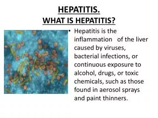
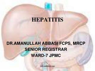
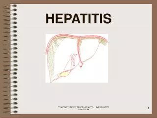
Hepatitis • Hepatitis is an inflammation of liver • It is usually caused by viral infections, toxic agents or drugs but may be an autoimmune response. • It is characterised by jaundice, abdominal pain, liver enlargement and sometimes fever. • It may be mild, or can be acute leading to fulminant hepatitis. Others, usually viral or alcoholic are chronic, and can lead to cirrhosis and liver cancer.
Characteristics of hepatitis viruses • F = faecal-oral route • P = parenteral
Hepatitis A virus (HAV) • Hep A virus • Picornaviridae; linear ss RNA; size 7.5 kb • Only one serotype, no antigenic cross-reactivity with HBV etc • Stable in 20% ether, acid pH1 for 2h, 60°C for 1h. • Destroyed by autoclaving 121°C for 20mins, boiling water 5 mins, dry heat 180°C for 1h, UV radiation, chlorine, formalin. • Heating food >85°C for 1min or disinfecting surfaces with sodium hypochlorite inactivate HAV. • HAV is detected in stool.
Hepatitis A • Faecal-oral route. • Spread in families, institutions, day care centers, neonatal intesive care units and among military troops. • An indication of poor sanitation and poor personal hygiene.
Identification • HAV – in stool and liver preparations • Immune EM • Measure Ab in serum • PCR • Tissue culture – no cytopathic effects are apparent. • Most cases will have complete recovery.
Hepatitis B virus (HBV) • Hepadnavirus • HBV establishes chronic infections esp in infants and a major factor of eventual hepatocellular carcinoma. • HBsAg – stability does not always conincide with that of the infectious agent. Both are stable at -20°C for over 20 yrs and stable to repeated freezing and thawing. • HBV but not HBsAg is sensitive to 100°C for 1 min or 60°C for 10hrs. HBsAg is stable at pH2.4 for <6hr. • HBsAg is not destroyed by UB irradiation of plasma or other blood products.
Hepatitis B • Other viruses, such as Yellow Fever, Epstein- Barr (EBV) and cytomegalovirus (CMV) as well as parasites and bacteria, can cause hepatitis as a secondary effect. • HBV is worldwide in distribution. • HBsAg can be detected in saliva, nasopharyngeal washings, semen. • Health care personnel (medical/dental surgeons, nurses, physicians) have higher risk and prevalence of detectable HBsAg or anti-HBs than those who have no occupational exposure to patients or blood products. They become apparently healthy HBsAg carriers.
Hep B Antigens • Hepatitis B DNA (HBV DNA) Immediate detection in bloodstream after initial infection ~1 week after infection. It maybe that the level of HBV DNA may indicate how fast the virus is replicating – using very sensitive and expensive PCR. It is generally only used research purposes, however in can be used to confirm the presence of hepatitis B and or measure viral load for viral mutants that do not produce the "e" and/or normal surface antigens. • Hepatitis B DNA polymerase. (HBV DNA Polymerase, DNAp) This enzyme can be detected in the bloodstream soon after initial infection by HBV at about the same time as HBV DNA ~ 1 week. Tests generally only used as indicators of disease progression, suitability for therapy and research purposes.
Hep B Antigens • Hepatitis B Core protein. (HBcAg) • Not detectable in the bloodstream, however it can be detected in the sample of liver cells taken after a liver biopsy. Generally the HBc proteins link together to form the hepatitis B core that encapsulate HBV DNA and DNA Polymerase. • Hepatitis B Surface protein(s). (HBsAg) • Outer surface coat composed of hepatitis B surface proteins is produced in larger quantities than required for the virus to reproduce. In some cases these particles encapsulate a core particle and produce a complete, and infectious, virus particle that enters the blood stream and can infect other liver cells. It does however take a while for these proteins to appear ~1 to 6 weeks before symptoms occur HBsAg appears. A positive test for the presence of hepatitis B surface protein (HBsAg), is the standard currently taken to indicate current infection with hepatitis B. If HBsAg is present for more than 6 months this is generally taken to indicate chronic infection. Majority of chronic HBV individuals are asymptomatic; may or may not have biochemical and histological evidence of liver disease. Chronic carriers are at high risk of hepatocellular carcinoma.
Hep B Antigens • HBe Protein. (HBeAg or 'e' antigen) • The Hepatitis 'e' antigen (HBeAg) is a peptide and representing the viraemic stage of hepatitis B virus, this in turn leads to the person being much more infectious and at a greater risk of progression to liver disease. The exact function of this non-structural protein is unknown, however it is thought that HBe may be influential in suppressing the immune systems response to HBV infection(?). HBeAg is generally detectable at the same time as HBsAg and disappears before HBsAg disappears. The presence of HBeAg in chronic infection is generally taken to indicate that HBV is actively reproducing and there is a higher probability of liver damage. In acute infection HBeAg is generally only transiently present. • However mutant strains of HBV exist that replicate without producing HBeAg. • In many cases infection with these mutant strains is more aggressive than HBe producing strains(?). • The presence of HBeAb is generally taken to be a good sign and indicates a favorable prognosis.
HBV vs Human • Majority of people will recover from Hepatitis B and around half of these are asymptomatic. Recovery means that no HBsAg is found in the blood and the Hepatitis B Antibody (HBsAb) is present. HBsAb usually persists for life after recovery. • These are generally the last antibodies to appear. HBsAb can neutralise the hepatitis B virus and there appearance taken as an indicator that an initial infection has been defeated. • Antibodies to HBc (HBcAb, Anti-HBc). • The first detectable antibody to appear around 8 weeks after infection with HBV are antibodies to the HBV core protein. The initial antibodies are of class IgM and IgG and generally appear after the appearance of HBsAg but often before ALT elevations. These antibodies to HBcAg do not neutralise the virus. HBcAb's persist in serum after an infection with HBV and these are predominantly of type IgG. The presence of a strong IgM HBcAb's is indicative of acute phase infection. The presence of IgG HBcAb's with no IgM HBcAb's antibody may be present in chronic and resolved cases of hepatitis B infection, this has been used to determine if the presence of HBsAb's was due to vaccination or by previous exposure to the live virus.
Detecting HBV • ALT alanine aminotransferase and AST (aspartate aminotransferase). • ALT and AST are enzymes produced in liver cells that can be detected in the blood stream. The normal range for ALT is between 0-40. When liver cells are damaged these enzymes are released and elevated levels are detected in serum. The value of ALT in the blood stream is generally taken to be an indicator of the damage that hepatitis causing to liver cells. However damage may be occurring with little or no elevation of ALT (this is especially true for hepatitis C and people with end stage liver disease). • ALT and AST and other substances are measured when a liver function test. However other drugs and especially alcohol can elevate these readings artificially.
Pathology • Acute hep B is characterised by spradically abnormal aminotransferase values and hepatomegaly. • Histologically, lobular architecture is preserved, portal inflammation and pale hepatocytes (cobblestone arrangement); and slight to absent fibrosis.
Chronic viral hepatitis type B frequently leads to a macronodular cirrhosis in non-allografted livers.
Hepatitis C virus • A positive-strand RNA virus; from family Flaviviridae • Hepatitis C is usually clinically mild, with minimal to moderate elevation of liver enzymes. • Despite the mild disease, 70-90% of cases progress to chronic liver disease. • Diseases in chronic HCV infections inc mixed cryoglobulinaemia and glomerulonephritis.
Hepatitis D virus • HDV infects all age groups – ppl with multiple transfusions, IV drug users, and their close contacts are at high risk. • HDV can be transmitted in a similar ways as HBV (though not sexually), and perinatally. • Infection is HBV-dependent, as HBV provides an HBsAg envelope for HDV. In some HBV infections, delta-Ag (HDAg) and anti-delta were detected. In blood, HCV contains HDAg surrounded by an HBsAg envelope. • Genome of HDV consists ss circular and negative-sense RNA; 1.7kbp (smallest).
Hepatitis E virus • Transmitted enterically; epidemic in developing countries where water supplies are sometimes faecally contaminated. • Viral genome is positive-sense ss RNA; 7.6 kbp in size.
Clinical findings • Not possible to make clinical distinction among cases caused by the hepatitis viruses. • Hepatitis may occur as complication of leptospirosis, syphilis, tuberculosis, all of which can be treated with drug therapy.
Treatment • Treatment of patients with hepatitis is supportive and allowing hepatocellular damage to resolve and repair itself. • Only HBV and HCV have specific treatments. • Recombinant interferon-α, lamivudine (reverse transcriptase inhibitor) reduces HBV DNA levels. • Orthotopic liver transplantation for chronic hepatitis B and C end-stage liver damage. However risk of reinfection on graft is at least 80% with HBV and 50% with HCV.
Prevention and Control • 1. Standard precautions when handling blood, body fluids and materials contaminated with hepatitis viruses – gloves, masks and eye protection, discard needles. • 2. HAV – formalin-inactivated HAV vaccines and reasonable hygiene. • 3. HBV - provides protection against infection with HBV by producing immunity or antibodies to the surface protein or outer coat of the virus, HBsAg. The first vaccine was produced by purifying this surface protein from the plasma of chronically infected persons. Subsequently, this surface protein was produced in yeast by recombinant DNA technology. However, plasma-derived vaccines continue to be used widely throughout the world. • 4. HCV – no vaccine.
Picornaviruses 19 Muharram 1428 7th February 2007 SBM 2044
Picornaviruses • PicoRNAvirus = small RNA virus • Include 2 main groups: • Enteroviruses – transient inhabitants of human alimentary tract, may be isolated from throat or lower intestine. • Rhinoviruses – isolated chiefly from nose and throat. • Cause diseases ranging from severe paralysis to aseptic meningitis, myocarditis. The most serious disease by enterovirus is poliomyelitis. • Picornaviruses replication occurs in cytoplasm.
Properties and Classification • Genome: ss RNA, linear, + sense, size is 7.2kb (human Rhinovirus) to 8.4kb (Aphthovirus). • No envelope, consists of 4 major polypeptides (surface VP1 and VP3 for Ab-binding sites; and internal VP4 is associated with viral RNA). • Enteroviruses are stable at acid pH (3-5) up to 3hr, whereas rhinoviruses are acid-labile.
I. Enterovirus Group • Poliomyelitis – acute infectious disease that affects the central nervous system. Destruction of motor neurons in spinal cord results in flaccid paralysis. Most infections are subclinical.
Pathogenesis and Pathology • Mouth is the portal entry of the virus, primary multiplication in oropharynx or intestine. • Virus is regularly present in throat and in the stools even before the onset of illness, or for many weeks even with a high Ab levels in blood. • It is believed that the virus multiplies in tonsils, LN of neck, Peyer’s patches, and circulating in the blood to infect CNS.
Clinical Findings • Abortive poliomyelitis – mild form of illness with fever, malaise, drowsiness, headache. Recovery in a few days. • Nonparalytic poliomyelitis (aseptic meningitis) – symptoms as above plus stiffness and pain in the back and neck; last 2-10 days. • Paralytic poliomyelitis – flaccid paralysis from lower motor neuron damage. Recovery within 6mths. • Progressive postpoliomyelitis muscle atrophy – reappearance of paralysis and muscle wasting decades after their paralytic poliomyelitis, a result of physiologic and aging changes in patients burdened by loss of neuromuscular functions.
Diagnosis • Virus recovered from throat swabs taken soon after onset of illness, stool samples. Uncommonly from the cerebrospinal fluid. • Identified by neutralisation with specific antiserum.
Prevention and Control • Periodic booster to maintain immunity. • Formalinised vaccine (Salk) is killed-virus vaccine, which induces humoral Abs but not local intestinal immunity so virus is still able to multiply in the gut. • Oral vaccines contain live attenuated virus (Sabin). This vaccine produces IgM, IgG and also secretory IgA Abs in intestine.
Coxsackieviruses • Produce diseases in humans such as: herpangina, hand-foot-and-mouth disease, acute haermorrhagic conjunctivitis by type A; and epidemic myalgia, myocarditis, pericarditis and generalised diseases in infants by type B. • Monkeys are mostly susceptible.
Echoviruses • Echoviruses = enteric cytopathogenic human orphan viruses. • Viruses may be recovered from throat and stools, similar to that of other enteroviruses. • Associated diseases are aseptic meningitis, rashes (“Boston exanthem disease”) which are common in young children, muscle weakness and spasm.
II. Rhinovirus Group • Are common cold viruses, cause upper resp T infections. • Can be found in nasal secretions within 2-4 days after exposure. Direct correlations between amount of virus and severity of illness. • Replication is limited to surface epithelium of nasal mucosa. • Immunity: Neutralising Ab develops in serum and secretions of most persons. Interferon may play a role in recorery – a 5-day course of intranasal IFN-α is an effective prevention within a family.
Foot-and-mouth Disease • Aphthovirus of Cattle • The disease in animals is highly contagious in the early stages of infection when viraemia is present when vesicles in the mouth and on the feet rupture and liberate large amount of virus. Excreted materials remain infectious for long periods. • Formalin-treated vaccines do not produce long-lasting immunity.
Reoviruses & Rotaviruses 19 Muharram 1428 7th February 2007 SBM 2044
General properties • Common disease is acute gastroenteritis. • Ds RNA, linear, icosahedral with double capsid shell. • No envelope, have 9 structural proteins with core contains several enzymes. • Reoviruses are usually stable to heat, pH 3-9 and to lipid solvents. • Replications occur in cytoplasm • Rotavirusesare major cause of infantile diarrhoea. • Reoviruses are good models for molecular studies of viral pathogenesis.
Reovirus Replication • Receptorsare viral haemagglutinin (α1 protein), a minor component of outer capsid. • Afterattachment and penetration, uncoating of virus occurs in lysosomes in cytoplasm. Only the outer shell of virus is removed, and core-associated RNA transcriptase is activated. • Gene expression : Reovirus cores contain all enzymes necs for transcribing, capping and extruding the mRNAs from the core, leaving the ds RNA genome inside.once extruded from the core, the mRNAs are translated into primary gene products. • Replication: viral replicase is responsible for synthesis of negative-sense strands to form ds genome segments. • Assembly of the correct complement segment is of unknown mechanism. May be self-assemble ?
Rotaviruses • Major cause of diarrhoeal illness in infants and young animals including calves and piglets. • Similar morphology and replication as Reovirus. • There are 5 groups (AE) based on antigenic epitopes of the internal structural protein VP6, with Group A is the most frequent human pathogen. • Outer capsids VP4 and VP7 are important neutralising antigens.
Pathogenesis • Rotaviruses infect cells in the villi of the small intestine (gastric and colonic mucosa are spared). • Multiply in cytoplasm of enterocytes and damage their transport mechanisms. • NSP4- a viral enterotoxin induces secretion by triggering a signal transduction pathway. Damaged cells may slough into lument and released as stools. • Diarrhoea may be due to impaired sodium and glucose absorption as damaged cells on villi are replaced by non-absorbing immature crypt cells. It may take 3-8 wks for normal function to be restored.