Download
1 / 56
570 likes | 742 Views
Central Nervous System Chapter 13 – Lecture Notes. to accompany Anatomy and Physiology: From Science to Life textbook by Gail Jenkins, Christopher Kemnitz, Gerard Tortora. Chapter Overview. 13.1 Central Nervous System 13.2 Protection and Nourishment of the CNS 13.3 Cerebrum

E N D
Central Nervous SystemChapter 13 – Lecture Notes to accompany Anatomy and Physiology: From Science to Life textbook by Gail Jenkins, Christopher Kemnitz, Gerard Tortora
Chapter Overview 13.1 Central Nervous System 13.2 Protection and Nourishment of the CNS 13.3 Cerebrum 13.4 Limbic System 13.5 Signal Processing in the Cerebrum 13.6 Diencephalon 13.7 Brain Stem 13.8 Cerebellum 13.9 Spinal Cord 13.10 Propagation of Impulses
Essential Terms Central Nervous System (CNS) • brain and spinal cord • control center for • thoughts • emotions • creativity • wisdom • memories • activities • behaviors Tract • bundle of axons
Introduction • CNS made up of ~100 billion neurons • Adult brain mass of ~1300g (3 lbs) • Spinal cord • mediates rapid reactions • reflexes • pathway for sensory nerve impulses to brain • pathway for motor nerve impulses from brain
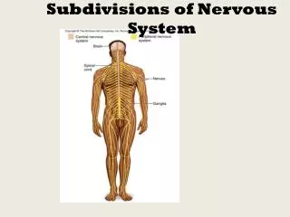
CNS Brain • cerebrum • cerebral hemispheres • diencephalon • brain stem • cerebellum Spinal Cord • medulla oblongata to superior edge of L2 Protection of CNS • two types of connective tissues • bony skull • cranial and spinal meninges • cushion of cerebrospinal fluid
Skeletal Protection • Brain is located in cranial cavity of skull • Spinal cord is located within vertebral canal of vertebral column • vertebral foramina of vertebrae stacked one on top of one another form the vertebral canal
Meninges • three connective tissue coverings that encircle brain and spinal cord • cranial meninges • spinal meninges • superficial to deep • dura mater • arachnoid mater • pia mater
Dura Mater of Brain • most superficial adheres to periosteum of cranial bones • strongest menix • extensions separate portions of brain • falx cerebri • two hemispheres of cerebrum • falx cerebelli • two hemispheres of cerebellum • tentorium cerebelli • separates cerebrum from cerebellum
Dura Mater of Spinal Cord • between dura mater and all of vertebral canal • epidural space • cushion of fat • dura mater tissue • sinuses that act as collection points for interstitial fluid and blood leaving brain • return interstitial fluid and blood to internal jugular veins of neck
Arachnoid Mater • avascular • collagen fibers • some elastic fibers • surrounds both brain and spinal cord • subdural space • thin space between dura mater and arachnoid matter • contains interstitial fluid
Pia Mater • innermost membrane • tightly adheres to surface of CNS • interlacing bundles of collagen fibers • some fine elastic fibers • surrounds both brain and spinal cord • subarachnoid space • thin space between arachnoid mater and pia matter • contains cerebrospinal fluid • also covers surface blood vessels of CNS
Meninges and Spinal Nerves • All three • cover spinal nerves • up to point of exit from spinal column • through intervertebral foramina
Denticulate Ligaments • suspend spinal cord in middle of dural sheath • membranous extensions of pia mater • project laterally and fuse with • arachnoid mater and • inner surface of dura mater • between anterior and posterior nerve roots of spinal nerves on either side • protect spinal cord against shock and sudden displacement
Blood Flow to CNS • to brain via • internal carotid and vertebral arteries • flows into dural sinuses • empties into internal jugular veins • to spinal cord via • posterior intercostal and lumbar arteries • empties into posterior intercostal and lumbar veins
Blood Flow to Brain • Brain at rest uses 20% of oxygen and glucose • even though only 2% of mass of adult • Neurons synthesize ATP almost exclusively from glucose • when activity increases in a particular region, blood flow to that area also increases
Blood Flow to Brain • decreased blood flow to brain • short time can cause unconsciousness • 1 to 2 minutes impairs neuronal function • 4 minutes causes permanent injury • virtually no glucose stored in the brain • low blood glucose to brain can cause • mental confusion • dizziness • convulsions • loss of consciousness
Blood Flow to Brain • Blocked blood flow to brain • arterial blockage can damage brain • CVA cerebrovascular accident • stroke • most common brain disorder • affect 500,000 people per year in US • 1/3 leading cause of death
Blood Brain Barrier Physiology • protects CNS from harmful • substances • pathogens • prevents passage from blood into interstitial fluid of neural tissue • water soluble substances usually pass by active transport • others pass slowly • lipid soluble substances pass readily
Blood Brain Barrier Anatomy • cerebral arteries divide quickly into capillaries • tight junctions seal together endothelial cells of CNS capillaries • capillaries also surrounded by thick basement membrane • astrocyte processes press against capillaries • selectively pass some substances and inhibit others
Cerebrospinal Fluid (CSF) • 80-150 ml volume • clear colorless liquid • protects and nourishes brain & spinal cord • protects • against chemical and physical injuries • acting as shock absorber on which brain floats • nourishes by carrying • oxygen • glucose • other chemicals • continuously circulates through cavities in and around CNS in subarachnoid space
Cerebrospinal Fluid (CSF) Contributes to homeostasis in three ways: • mechanical protection • shock absorber • chemical protection • circulation
Formation of CSF • CSF fills ventricles • lateral ventricles • located in each hemisphere of cerebrum • separated by septum pellucidum • third ventricle • fourth ventricle • CSF produced in choroid plexuses • capillaries in walls of ventricles • covered by ependymal cells that form CSF from blood plasma by filtration and secretion
Circulation of CSF • Cilia on ependymal cells assist with flow • from lateral ventricles • through interventricular foramina • to third ventricle • then through cerebral aqueduct • into fourth ventricle • enters subarachnoid space through • median aperture • pair of lateral apertures • reabsorbed into blood • arachnoid villi
Cerebrum • Seat of intelligence • interprets sensory impulses • controls muscular movements • functions in emotional and intellectual processes • Cerebral Cortex • gray matter on outside • receives & integrates incoming & outgoing information • White matter on inside • white is myelination • Gray matter nuclei deep within white matter
Cerebral Cortex • enlarges faster during embryonic development than white matter • rolls and folds forming • gyri (singular = gyrus) • bulges or folds • fissures • deep grooves • longitudinal fissure separates cerebrum into left and right hemispheres • connected internally by corpus callosum • sulci (singular = sulcus) • shallow fissures
Cerebral White Matter • has tracts • myelinated and unmyelinated axons • communicate between regions of CNS • three types • association tracts • between gyri in same hemisphere • commissural tracts • from gyri in one hemisphere to corresponding gyri in other hemisphere • projection tracts • from cerebrum to lower parts of CNS
Basal Nuclei • mass of cell bodies • two are side by side just lateral to thalamus • globus pallidus and putamen • third is caudate nucleus • large “head” connected to smaller “tail” by long comma-shaped “body” • receive input from cerebral cortex • provide output to motor portions • control subconscious contractions of skeletal muscles