Download
1 / 62
620 likes | 649 Views
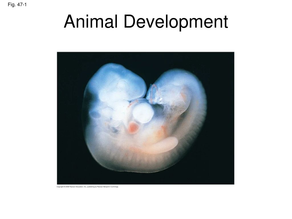
Fig. 47-1. Animal Development. 1 mm. Development is determined by the zygote’s genome and molecules in the egg called cytoplasmic determinants Cell differentiation is the specialization of cells in structure and function Morphogenesis is the process by which an animal takes shape.

E N D
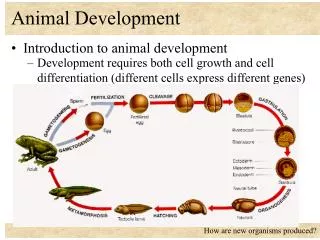
Fig. 47-1 Animal Development 1 mm
Development is determined by the zygote’s genome and molecules in the egg called cytoplasmic determinants • Cell differentiation is the specialization of cells in structure and function • Morphogenesis is the process by which an animal takes shape
After fertilization, embryonic development proceeds through cleavage, gastrulation, and organogenesis • Important events regulating development occur during fertilization and the three stages that build the animal’s body • Cleavage: cell division creates a hollow ball of cells called a blastula • Gastrulation: cells are rearranged into a three-layered gastrula • Organogenesis: the three layers interact and move to give rise to organs
Fertilization • Fertilization brings the haploid nuclei of sperm and egg together, forming a diploid zygote • The sperm’s contact with the egg’s surface initiates metabolic reactions in the egg that trigger the onset of embryonic development
The Acrosomal Reaction • The acrosomal reaction is triggered when the sperm meets the egg • The acrosome at the tip of the sperm releases hydrolytic enzymes that digest material surrounding the egg
Fig. 47-3-5 Sperm plasmamembrane Spermnucleus Fertilizationenvelope Acrosomalprocess Basal body(centriole) Actinfilament Spermhead Corticalgranule Fusedplasmamembranes Perivitellinespace Hydrolytic enzymes Acrosome Jelly coat Vitelline layer Sperm-bindingreceptors Egg plasmamembrane EGG CYTOPLASM
Gamete contact and/or fusion depolarizes the egg cell membrane and sets up a fast block to polyspermy
The Cortical Reaction • Fusion of egg and sperm also initiates the cortical reaction • This reaction induces a rise in Ca2+ that stimulates cortical granules to release their contents outside the egg • These changes cause formation of a fertilization envelope that functions as a slow block to polyspermy
Activation of the Egg • The sharp rise in Ca2+ in the egg’s cytosol increases the rates of cellular respiration and protein synthesis by the egg cell • With these rapid changes in metabolism, the egg is said to be activated • The sperm nucleus merges with the egg nucleus and cell division begins
Fertilization in Mammals • Fertilization in mammals and other terrestrial animals is internal • In mammalian fertilization, the cortical reaction modifies the zona pellucida,the extracellular matrix of the egg,as a slow block to polyspermy
Fig. 47-5 Zona pellucida Follicle cell Cortical granules Spermnucleus Spermbasal body
In mammals the first cell division occurs 12–36 hours after sperm binding • The diploid nucleus forms after this first division of the zygote
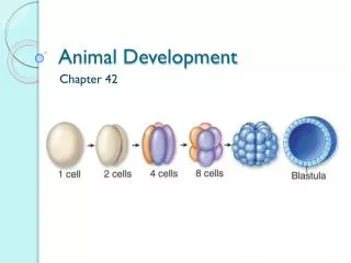
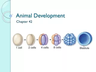
Cleavage • Fertilization is followed by cleavage, a period of rapid cell division without growth • Cleavage partitions the cytoplasm of one large cell into many smaller cells called blastomeres • The blastula is a ball of cells with a fluid-filled cavity called a blastocoel
Fig. 47-6 (a) Fertilized egg (b) Four-cell stage (c) Early blastula (d) Later blastula
The eggs and zygotes of many animals, except mammals, have a definite polarity • The polarity is defined by distribution of yolk (stored nutrients) • The vegetal pole has more yolk; the animal pole has less yolk
The three body axes are established by the egg’s polarity and by a cortical rotation following binding of the sperm • Cortical rotation exposes a gray crescent opposite to the point of sperm entry
Fig. 47-7 Dorsal Right Anterior Posterior Left Ventral (a) The three axes of the fully developed embryo Animal pole Firstcleavage Pigmentedcortex Point ofspermnucleusentry Animalhemisphere Futuredorsalside Vegetalhemisphere Graycrescent Vegetal pole (b) Establishing the axes
Cleavage planes usually follow a pattern that is relative to the zygote’s animal and vegetal poles
Fig. 47-8-6 0.25 mm 0.25 mm Animal pole Blastocoel Vegetalpole Zygote 2-cellstageforming 4-cellstageforming 8-cellstage Blastula(crosssection)
Cell division is slowed by yolk • Holoblastic cleavage, complete division of the egg, occurs in species whose eggs have little or moderate amounts of yolk, such as sea urchins and frogs
Meroblastic cleavage, incomplete division of the egg, occurs in species with yolk-rich eggs, such as reptiles and birds
Gastrulation • Gastrulation rearranges the cells of a blastula into a three-layered embryo, called a gastrula, which has a primitive gut
The three layers produced by gastrulation are called embryonic germ layers • The ectoderm forms the outer layer • The endoderm lines the digestive tract • The mesoderm partly fills the space between the endoderm and ectoderm
Gastrulation in the sea urchin embryo • The blastula consists of a single layer of cells surrounding the blastocoel • Mesenchyme cells migrate from the vegetal pole into the blastocoel • The vegetal plate forms from the remaining cells of the vegetal pole and buckles inward through invagination
Gastrulation in the sea urchin embryo • The newly formed cavity is called the archenteron • This opens through the blastopore, which will become the anus
Fig. 47-9-6 Key Future ectoderm Future mesoderm Future endoderm Archenteron Blastocoel Filopodiapullingarchenterontip Animalpole Blastocoel Archenteron Blastocoel Blastopore Mesenchymecells Ectoderm Vegetalplate Vegetalpole Mouth Mesenchymecells Mesenchyme(mesodermforms futureskeleton) Digestive tube (endoderm) Blastopore 50 µm Anus (from blastopore)
Gastrulation in the chick • The embryo forms from a blastoderm and sits on top of a large yolk mass • During gastrulation, the upper layer of the blastoderm (epiblast) moves toward the midline of the blastoderm and then into the embryo toward the yolk
The midline thickens and is called the primitive streak • The movement of different epiblast cells gives rise to the endoderm, mesoderm, and ectoderm
Fig. 47-11 Dorsal Fertilized egg Primitive streak Anterior Embryo Left Right Yolk Posterior Ventral Primitive streak Epiblast Futureectoderm Blastocoel Endoderm Migratingcells(mesoderm) Hypoblast YOLK
Organogenesis • During organogenesis, various regions of the germ layers develop into rudimentary organs • The frog is used as a model for organogenesis
Early in vertebrate organogenesis, the notochord forms from mesoderm, and the neural plate forms from ectoderm
Fig. 47-12 Eye Somites Tail bud Neural folds Neural plate Neuralfold SEM 1 mm 1 mm Neural crestcells Neural tube Neuralfold Neural plate Notochord Coelom Neural crestcells Somite Notochord Ectoderm Archenteron(digestivecavity) Outer layerof ectoderm Mesoderm Endoderm Neural crestcells (c) Somites Archenteron (a) Neural plate formation Neural tube (b) Neural tube formation
Fig. 47-12a Neural plate Neuralfold 1 mm Neural folds Notochord Ectoderm Mesoderm Endoderm Archenteron (a) Neural plate formation
The neural plate soon curves inward, forming the neural tube • The neural tube will become the central nervous system (brain and spinal cord)
Fig. 47-12b-1 Neuralfold Neural plate (b) Neural tube formation
Fig. 47-12b-2 (b) Neural tube formation
Fig. 47-12b-3 Neural crestcells (b) Neural tube formation
Fig. 47-12b-4 Outer layerof ectoderm Neural crestcells Neural tube (b) Neural tube formation
Neural crest cells develop along the neural tube of vertebrates and form various parts of the embryo (nerves, parts of teeth, skull bones, and so on) • Mesoderm lateral to the notochord forms blocks called somites • Lateral to the somites, the mesoderm splits to form the coelom
Fig. 47-12c Neural crestcells Neural tube Somites Eye Tail bud Notochord Coelom Somite Archenteron(digestivecavity) SEM 1 mm (c) Somites
Organogenesis in the chick is quite similar to that in the frog
Fig. 47-13 Eye Neural tube Notochord Forebrain Somite Heart Coelom Archenteron Endoderm Lateral fold Mesoderm Bloodvessels Ectoderm Somites Yolk stalk Yolk sac These layersform extraembryonicmembranes Neural tube YOLK (a) Early organogenesis (b) Late organogenesis
Fig. 47-14 ECTODERM MESODERM ENDODERM NotochordSkeletal systemMuscular systemMuscular layer ofstomach and intestineExcretory systemCirculatory and lymphaticsystems Reproductive system(except germ cells) Dermis of skinLining of body cavityAdrenal cortex Epidermis of skin and itsderivatives (including sweatglands, hair follicles)Epithelial lining of mouthand anusCornea and lens of eyeNervous systemSensory receptors inepidermisAdrenal medullaTooth enamelEpithelium of pineal andpituitary glands Epithelial lining ofdigestive tractEpithelial lining ofrespiratory systemLining of urethra, urinarybladder, and reproductivesystemLiverPancreasThymusThyroid and parathyroidglands
Developmental Adaptations of Amniotes • Embryos of birds, other reptiles, and mammals develop in a fluid-filled sac in a shell or the uterus • Organisms with these adaptations are called amniotes
During amniote development, four extraembryonic membranes form around the embryo: • The chorion functions in gas exchange • The amnion encloses the amniotic fluid • The yolk sac encloses the yolk • The allantois disposes of waste products and contributes to gas exchange
Fig. 47-15 Amnion Allantois Embryo Amnioticcavitywithamniotic fluid Albumen Shell Yolk (nutrients) Chorion Yolk sac
Mammalian Development • The eggs of placental mammals • Are small and store few nutrients • Exhibit holoblastic cleavage • Show no obvious polarity • Gastrulation and organogenesis resemble the processes in birds and other reptiles • Early cleavage is relatively slow in humans and other mammals
At completion of cleavage, the blastocyst forms • A group of cells called the inner cell mass develops into the embryo and forms the extraembryonic membranes • The trophoblast, the outer epithelium of the blastocyst, initiates implantation in the uterus, and the inner cell mass of the blastocyst forms a flat disk of cells • As implantation is completed, gastrulation begins
Fig. 47-16-1 Endometrialepithelium(uterine lining) Uterus Inner cell mass Trophoblast Blastocoel
The epiblast cells invaginate through a primitive streak to form mesoderm and endoderm • The placenta is formed from the trophoblast, mesodermal cells from the epiblast, and adjacent endometrial tissue • The placenta allows for the exchange of materials between the mother and embryo • By the end of gastrulation, the embryonic germ layers have formed