Download
1 / 64
660 likes | 816 Views
Animal Development. Chapter 46 & 47. Fig. 46-6. Vocabulary. Zygote: Single diploid cell Fertilization: Sperm & egg combine Ovulation: Egg is released from the ovary Spermatogenesis: Formation of sperm Oogenesis: Formation of the egg. Anatomy. Anatomy. Oviduct. Ovary. Uterus.

E N D
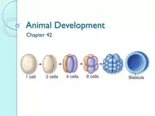
Animal Development Chapter 46 & 47
Vocabulary • Zygote: • Single diploid cell • Fertilization: • Sperm & egg combine • Ovulation: • Egg is released from the ovary • Spermatogenesis: • Formation of sperm • Oogenesis: • Formation of the egg
Anatomy Oviduct Ovary Uterus (Urinary bladder) (Pubic bone) (Rectum) Cervix Urethra Shaft Vagina Clitoris Glans Prepuce Labia minora Labia majora Vaginal opening
Fig. 46-10b Oviduct Ovaries Follicles Corpus luteum Uterine wall Uterus Endometrium Cervix Vagina
Female • Ovaries (at birth) • Contain approximately 1 million follicles • Primary oocyte • Started meiosis • Stopped at prophase I (diploid)
Female • FSH • Stimulates production of a follicle • One follicle goes through Meiosis I • Two daughter cells • Secondary oocyte (starts Meiosis II) • Polar body (disintegrates)
Female • LH stimulates ovary • Secondary oocyte leaves ovary • Ovulation • Fertilized then completes Meiosis II • Ovum • Polar body • Travels fallopian tube • Implants in uterus (approx. 5-6 days)
Fig. 46-12g In embryo Primordial germ cell Mitotic divisions 2n Oogonium Mitotic divisions Primary oocyte(present at birth), arrestedin prophase of meiosis I 2n Completion of meiosis Iand onset of meiosis II Firstpolarbody n n Secondary oocyte,arrested at metaphase of meiosis II Ovulation, sperm entry Completion of meiosis II Secondpolarbody n Fertilized egg n
Menstrual cycle • GnRH • FSH & LH released • Stimulates follicle • Estradiol released • Follicle released (ovulation) • LH increases
Menstrual cycle • Corpus luteum releases progesterone & estridiol • Corpus luteum disintegrates • Lining sheds • Endometriosis: • Uterine lining in abdomen
Fig. 46-11b (Urinarybladder) (Urinaryduct) Seminal vesicle (Rectum) (Pubic bone) Vas deferens Erectiletissue Ejaculatory duct Prostate gland Urethra Penis Bulbourethral gland Glans Vas deferens EpididymisTestisScrotum Prepuce
Male • Testes are in abdomen (at birth) • Descend into scrotum • Temperature of testes is cooler • Normal sperm production • Seminiferous tubules • Contain spermatogonia or germ cells
Male • Germ cells (diploid) • Mitosis • One undergoes meiosis • Produce 4 haploid sperm • Produce 100 to 200 million sperm a day • Continues for life
Fig. 46-12b Epididymis Seminiferous tubule Sertoli cellnucleus Spermatogonium Primary spermatocyte Testis Cross sectionof seminiferoustubule Secondary spermatocyte Spermatids(two stages) Sperm Lumen ofseminiferous tubule
Fig. 46-12c Primordial germ cell in embryo Mitotic divisions Spermatogonialstem cell 2n Mitotic divisions Spermatogonium 2n Mitotic divisions Primary spermatocyte 2n Meiosis I n n Secondary spermatocyte Meiosis II Earlyspermatid n n n n Differentiation (Sertolicells provide nutrients) Sperm n n n n
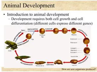
Embryonic development • Fertilization • Cleavage • Gastrulation • Neurulation • Organogenesis
Fertilization • 1. Penetration • Sperm digests cells surrounding egg • Contains glycoprotein enzymes • 2. Activation • Membrane changes • Prevents other sperm penetrating
Fertilization • 2. Activation • A. stimulates egg to complete division of Meiosis II • B. stimulates movement of cytoplasm to prepare for cell division of zygote • C. stimulates increase in protein synthesis
Fertilization • 3. Nuclei fusion • Sperm nucleus fuses with egg • Egg is not activated • Does not form zygote
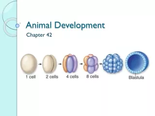
Cleavage • Rapid cell division • Blastomeres: • Smaller & smaller cells • No increase in volume of cytoplasm • Morula: • Tight mass of approximately 32 cells
Fig. 47-6 (a) Fertilized egg (b) Four-cell stage (c) Early blastula (d) Later blastula
Cleavage • Blastocyst (Blastula) • Hollow ball of approx. 500-2000 cells • Blastocyst cavity • Fluid filled • Different regions in blastocyst • Received differing amounts of cytoplasm • Affects further development
Cleavage • Trophoblast: • Outer layer of cells • Surround blastocyst (involved in placenta) • Inner cell mass: • Layer of dividing cells • At one end of Blastocyst • Becomes developing embryo
Fig. 47-8-6 0.25 mm 0.25 mm Animal pole Blastocoel Vegetalpole Zygote 2-cellstageforming 4-cellstageforming 8-cellstage Blastula(crosssection)
Cleavage • Implantation: • Blastocyst attaches to endometrium • 6 days after fertilization • Human chorionic gonadotropin (HCG) • Hormone released by trophoblast • Maintains corpus luteum
Fig. 47-16-2 Expandingregion oftrophoblast Maternalbloodvessel Epiblast Hypoblast Trophoblast
Gastrulation • Turning inward of cells into blastocyst • Forms germ layers • Ectoderm: • Epidermis/neural tissue • Mesoderm: • Muscle/skeletal/vasculature • Endoderm: • Gut lining, respiratory tract, liver
Fig. 47-14 ECTODERM MESODERM ENDODERM NotochordSkeletal systemMuscular systemMuscular layer ofstomach and intestineExcretory systemCirculatory and lymphaticsystems Reproductive system(except germ cells) Dermis of skinLining of body cavityAdrenal cortex Epidermis of skin and itsderivatives (including sweatglands, hair follicles)Epithelial lining of mouthand anusCornea and lens of eyeNervous systemSensory receptors inepidermisAdrenal medullaTooth enamelEpithelium of pineal andpituitary glands Epithelial lining ofdigestive tractEpithelial lining ofrespiratory systemLining of urethra, urinarybladder, and reproductivesystemLiverPancreasThymusThyroid and parathyroidglands
Gastrulation • Chorion: • Surrounds embryo • Gas exchange • Amnion: • Encloses the embryo • Protective amniotic fluid • Yolk sac: • Formation of blood cells
Gastrulation Amnion Chorion Ectoderm Mesoderm Endoderm Yolk sac Extraembryonicmesoderm Atlantois
Neurulation • Development of dorsal nerve cord • Notochord (spinal column) • Forms from mesoderm soon after gastrulation • Neural grove (spinal cord/brain) • Crease down the axis of the embryo • Neural tube (ectoderm) • Hollow cylinder
Neurulation • Neural crest • Cells pinch off from neural tube • Migrate to parts of embryo • Peripheral nerves, teeth, skull bones
Fig. 47-12b-4 Outer layerof ectoderm Neural crestcells Neural tube (b) Neural tube formation
Organogenesis • Below neural tube • Somitomeres: • Small sections of mesoderm • Somites • Smaller sections of mesoderm • Develop into muscles, vertebrae, connective tissues
Organogenesis • Mesoderm surrounds the endoderm • Separates into 2 layers • One lines the inner body wall • One lines the outside of the gut • Between layers becomes body cavity