Download
1 / 74
740 likes | 745 Views
Cancer. When good cells go bad. What is cancer?. Caner is defined as the continuous uncontrolled growth of cells. A tumor is a any abnormal proliferation of cells. Benign tumors stays confined to its original location

E N D
Cancer When good cells go bad
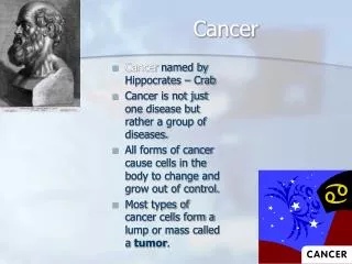
What is cancer? • Caner is defined as the continuous uncontrolled growth of cells. • A tumor is a any abnormal proliferation of cells. • Benign tumors stays confined to its original location • Malignant tumors are capable of invading surrounding tissue or invading the entire body • Tumors are classified as to their cell type • Tumors can arise from any cell type in the body
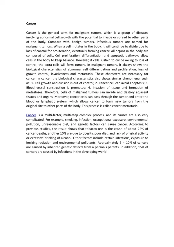
Cancer is an umbrella term covering a plethora of conditions characterized by unscheduled and uncontrolled cellular proliferation. • The causes of cancer are many and varied, and include genetic predisposition, environmental influences, infectious agents and aging. These transform normal cells into cancerous ones by derailing a wide spectrum of regulatory and downstream effector pathways. It is just this complexity that has hampered the development of effective and specific cancer therapies.
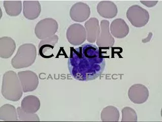
Cancer continued; three cancer types • Carcinomas; constitute 90% of cancers, are cancers of epithelial cells • Sarcomas; are rare and consist of tumors of connective tissues (connective tissue, muscle, bone etc.) • Leukemias and lymphomas; constitute 8% of tumors. Sometimes referred to as liquid tumors. Leukemias arise from blood forming cells and lymphomas arise from cells of the immune system (T and B cells).
Properties of cancer cells Cancer cells lack contact inhibition Normal cells show contact inhibition
Properties of cancer cells They keep growing And growing And growing And growing
Prevalence of Factors That Modify the Risk of Cancer in the United States. Emmons KM, Colditz GA. N Engl J Med 2017;376:986-990.
Cancer Fig 16.3 • Cells in culture and in vivo exhibit contact-inhibition • Cancer cells lack contact inhibition feedback mechanisms. Clumps or foci develop.
Cancer: Benign • Benign: localized and of small size • Cells that closely resemble, and may function, like normal cells • May be delineated by a fibrous (Basal lamina) capsule • Become problems due to sheer bulk or due to secretions (e.g. hormones)
Cancer : Malignant Malignant tumors: high rate of division, properties may vary compared to cells of origin. Most malignant cells become metastatic Invade surrounding tissue and establishment of secondary areas of growth: Metastasis
1. Growth Factor Receptor Increased numbers in 20 percent of breast cancers 2. Ras Protein Activated by mutations in 20 to 30 percent of cancers 3. Abl Kinase Activated by abnormal chromosomes in chronic myelogenous leukemia 4. Src Kinase Activated by mutations in 2 to 5 percent of cancers 5. p53 Protein Mutated or deleted in 50 percent of cancers ASSOCIATION WITH HUMAN CANCERS
B. Virology DNA tumor viruses- subvert cellular machinery for replication Adenovirus: Early: dedicated to replication of genome. Triggered by E1A Need host cell to be in S-phase, and E1A does the job Uses host cell factors to activate transcription of essential early viral genes. (late: viral capsid/packaging proteins) Immortalization characterized by increased S-phase entry-overcome a G1 block
What cellular proteins bind E1A and SV40 Large T?? (Harlow, Livingston) Objective: provide clues into the cellular pathway. What kinds of proteins co-IP’s with E1A? (anti E1A IP from 35S-cells) RB Family: p105, p107, p130 Cell Cycle: Cyclin A, CDK2 For E1A and Lg.T: LXCXE A B RB Model E1A neutralized RB growth arrest to enhance S-phase.
RB PATHWAY The Retinoblastoma Family: pRB, p107, p130 Focus mainly on RB (Merger of virology, genetics, and cell biology) A. Genetics/Tumor Suppressors The concept of tumor suppressor protein came from studies of retinoblastomas--tumors of the eye. Found loss of heterozygosity in a particular position in the chromosome.
When gene was cloned-p105-110 A. highly mutated in retinoblastomas B. many other tumors have mutations. Mutations in tumors: in a pocket region. Led to the idea that the normal function is the suppression of cell growth. Over expression leads to suppression of growth. Nuclear phosphoprotein
pRB Pathway Mitogenic Stimuli (e.g. GF, Ras) RB X E2F D-Cyclin CDK 4/6 DNA Pol Cyclin E, p19 DHFR, MYB E2F PPP P16 Ink4a RB Tumor Suppressor Genes RB, p16 Oncogenes Cyclin D1 From Sharpless and DePinho (1999) Current Opinions in Genetics and Dev. 9:22
Cancer Fig. 16.13 136371
Viral Oncogenes Induce Proliferation and Suppress Apoptosis G1 Adenovirus E1A HPV E7 SV40 Lg T RB S p53 APOPTOSIS Adenovirus E1B(55K) HPV E6 SV40 Lg T Adenovirus E1B (19K) (Bcl2-like)
p53 in apoptosis Following DNA damage, e.g. by radiation, p53 levels rise, and proliferating cells arrest in G1. This allows time for DNA repair prior to the next round of replication. This arrest is mediated by stimulation of expression of p21CIP1, the cyclin kinase inhibitor. Very high p53 levels, or susceptible cell types, e.g. lymphocytes, are triggered to undergo apoptosis. Bcl-2 acts between p53 and the caspase:
Apoptosis • Apoptosis is a tightly regulated form of cell death, also called the programmed cell death. Morphologically, it is characterized by chromatin condensation and cell shrinkage in the early stage. Then the nucleus and cytoplasm fragment, forming membrane-bound apoptotic bodies which can be engulfed by phagocytes. In contrast, cells undergo another form of cell death, necrosis, swell and rupture. The released intracellular contents can damage surrounding cells and often cause inflammation.