Download
1 / 70
770 likes | 1.15k Views
UNIT 4 DNA and Its Role in Heredity PART ONE: DNA Structure/Function. Hillis Textbook: Chapter9. GENETIC MATERIAL CRITERIA:. Scientists had criteria for DNA to be accepted as the genetic material, including that it: Be present in the cell nucleus and in chromosomes Doubles in the cell cycle

E N D
UNIT 4DNA and Its Role in HeredityPART ONE:DNA Structure/Function • Hillis Textbook: Chapter9
GENETIC MATERIAL CRITERIA: • Scientists had criteria for DNA to be accepted as the genetic material, including that it: • Be present in the cell nucleus and in chromosomes • Doubles in the cell cycle • Is twice as abundant in diploid cells • Has same the pattern of transmission as its genetic information
MIESCHER: • DNA was found in the nucleus by Miescher. • He isolated cell nuclei and treated them chemically. • A fibrous substance came out of the solution and he called it “nuclein”. • DNA was found in chromosomes using dyes that bind specifically to DNA.
GENETIC MATERIAL WAS FOUND: • Dividing cells were stained and passed through a flow cytometer, confirming two other predictions for DNA: • Nondividing cells have the same amount of nuclear DNA. • After meiosis, gametes have half the amount of DNA.
GENETIC MATERIAL WAS FOUND: • Chromosomes contain DNA, but also contain proteins, so scientists had to determine whether proteins carried genetic information. • Viruses, such as bacteriophages, contain DNA and a little protein. • When a virus infects a bacterium, it injects only its DNA into it, and changes the genetic program of the bacterium. • This provides further evidence for DNA, and not protein, as the genetic material.
Figure 9.2 Viral DNA and Not Protein Enters Host Cells (Part 2)
GENETIC MATERIAL WAS FOUND: • Bacterial Transformation experiments showed that DNA from one strain of bacteria could genetically transform (make) another strain.
GENETIC MATERIAL WAS FOUND: • After identifying DNA as the genetic material, scientists hoped to answer two questions about the structure: • How is DNA replicated between cell divisions? • How does it direct the synthesis of specific proteins?
DNA STRUCTURE: • DNA structure was discovered through the work of many scientists. • One crucial piece of evidence came from X-ray crystallography. • A purified substance can be made to form crystals; the pattern of diffraction of X rays passed through the crystallized substance shows position of atoms.
ROSALIND FRANKLIN: • Rosalind Franklin: • Prepared crystallographs from uniformly oriented DNA fibers—her images suggested a spiral model (helix)
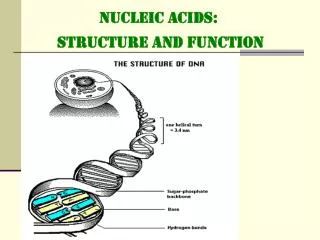
GENETIC MATERIAL: • Chemical composition also provided clues: • DNA is a polymer of nucleotides:deoxyribose sugar, a phosphate group, and a nitrogen-containing base. • The bases form the differences between nucleotides: • Purines: Pyrimidines: • adenine (A) cytosine (C) • guanine (G) thymine (T)
ERWIN CHARGAFF: • In 1950 Erwin Chargaff found that in the DNA from many different species: • Amount of A = amount of T • Amount of C = amount of G • Or, the abundance of purines = the abundance of pyrimidines • Chargaff’s rule.
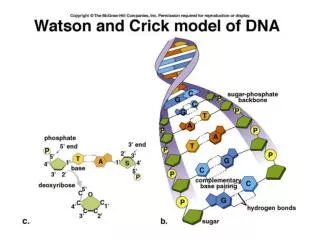
WATSON AND CRICK: • “Model building” is the assembly of 3-D models of possible molecular structures. • Francis Crick and James Watson used model building and combined all the knowledge of DNA to determine its structure. • Franklin’s X-ray crystallography convinced them the molecule was helical. • Modeling also showed that DNA strands are anti-parallel.
WATSON AND CRICK: • Watson and Crick suggested that: • Nucleotide bases are on the interior of the two strands, with a sugar-phosphate backbone on the outside. • Per Chargaff’s rule, a purine on one strand is paired with a pyrimidine on the other. • These base pairs (A-T and G-C) have the same width down the helix.
KEY FEATURES: • Four key features of DNA structure: • It is a double-stranded helix of uniform diameter. • It is right-handedand twists CLOCKWISE. • It is antiparallel– runs opposite directions. • Outer edges of nitrogenous bases are exposed in the major and minor grooves. • Major grooves – larger gap • Minor grooves – smaller gap
Figure 9.6 Base Pairs in DNA Can Interact with Other Molecules
FUNCTIONS OF DNA: • DNA has four important functions— • double-helical structure is essential: • 1. Storage of genetic information—millions of nucleotides; base sequence encodes huge amounts of information • 2. Precise replication during cell division by complementary base pairing • 3. Susceptibility to mutations—a change in information—possibly a simple alteration to a sequence • 4. Expression of the coded information as the phenotype—nucleotide sequence is transcribed into RNA and determines sequence of amino acids in proteins
UNIT 4DNA and Its Role in HeredityPART TWO:DNA Replication • Hillis Textbook: Chapter9
REPLICATION OF DNA: • Semiconservative replication means that each parental strand serves as a template for a new strand (separately). • Conservative replication would show that the intact parental DNA (both strands) serves as a template. • Evidence from radioactively-labeled strands supports semiconservative replication.
REPLICATION OF DNA: • Two steps in DNA replication: • 1.The double helix is unwound, making two template strands available for new base pairing. • 2.New nucleotides form base pairs with template strands and linked together by phosphodiester bonds. Template DNA is read in the 3′-to-5′ direction.
REPLICATION OF DNA: • During DNA synthesis, new nucleotides are added to the 3′ end of the new strand, which has a free hydroxyl group (—OH). • Deoxyribonucleoside triphosphates (dNTPs), or deoxyribonucleotides, are the building blocks—two of their phosphate groups are released and the third bonds to the 3′ end of the DNA chain.
Figure 9.7 Each New DNA Strand Grows by the Addition of Nucleotides to Its 3′ End
REPLICATION OF DNA: • STEP ONE: • DNA replication begins with the binding of a large protein complex—the pre-replication complex—to a specific site on the DNA molecule. • The complex contains DNA polymerase, which catalyzes addition of nucleotides. • The complex binds to a region on the chromosome called the origin of replication (ori).
REPLICATION OF DNA: • STEP TWO: • When the pre-replication complex binds to ori, the DNA unwinds and replication proceeds in two directions. • The replication fork is the site where DNA unwinds to expose bases. • Eukaryotic chromosomes are linear and have multiple origins of replication, which speed up replication.
REPLICATION OF DNA: • STEP THREE: • DNA replication begins with a short primer—a starter strand. (ACTUALLY RNA) • The primer is complementary to the DNA template. • Primase—an enzyme—synthesizes DNA primer one nucleotide at a time. • DNA polymerase adds nucleotides to the 3′ end.
REPLICATION OF DNA: • DNA polymerases are larger than their substrates, the dNTPs, and the template DNA. • The enzyme is shaped like an open right hand—the “palm” brings the active site and the substrates into contact. • The“fingers”recognize the nucleotide bases.
Figure 9.10 DNA Polymerase Binds to the Template Strand (Part 2)
REPLICATION OF DNA: • STEP FOUR: • A single replication fork opens up in one direction. • The two DNA strands are antiparallel—the 3′ end of one strand is paired with the 5′ end of the other. • DNA replicates in a 5′-to-3′ direction on the lagging strand
REPLICATION OF DNA: • STEP FIVE: • One new strand, the leading strand, is oriented to grow at its 3′ end as the fork opens. • The lagging strand is oriented so that its exposed 3′ end gets farther from the fork. • Synthesis of the lagging strand occurs in small, discontinuous stretches—Okazaki fragments.
REPLICATION OF DNA: • STEP SIX: • Each Okazaki fragment requires its own primer, synthesized by the primase. • DNA polymerase adds nucleotides to the 3′ end, until reaching the primer of the previous fragment. • A different DNA polymerase then replaces the primer with DNA. • The final phosphodiester linkage between fragments is catalyzed by DNA ligase.
Figure 9.12 The Lagging Strand Story (Part 3) DNA polymerase works very fast: It is processive—it catalyzes many sequential polymerization reactions each time it binds to DNA
REPLICATION OF DNA: • STEP SEVEN: • Okazaki fragments are added to RNA primers to replicate the lagging strand. • When the last primer is removed no DNA synthesis occurs because there is no 3′ end to extend—a single-stranded bit of DNA is left at each end. • These are cut after replication and the chromosome is slightly shortened after each cell division.
Concept 9.2 DNA Replicates Semiconservatively • Telomeres are repetitive sequences at the ends of eukaryotic chromosomes. • These repeats prevent the chromosome ends from being joined together by the DNA repair system. • Telomerase contains an RNA sequence—it acts as a template for telomeric DNA sequences. • Telomeric DNA is lost over time in most cells, but not in continuously dividing cells like bone marrow and gametes.
REPLICATION OF DNA: • DNA polymerases can make mistakes in replication, but most errors are repaired. • Cells have two major repair mechanisms: • Proofreading—as DNA polymerase adds nucleotides, it has a proofreading function and if bases are paired incorrectly, the nucleotide is removed. • Mismatch repair—after replication other proteins scan formismatched bases missed in proofreading, and replace them with correct ones.
Concept 9.2 DNA Replicates Semiconservatively • Copies of DNA sequences can be made by the polymerase chain reaction (PCR) technique, which uses: • A double-stranded DNA sample • Two short primers complementary to the ends of the sequence to be amplified • The four dNTPs • A DNA polymerase that works at high temperatures • Salts and a buffer to maintain pH
UNIT 4DNA and Its Role in HeredityPART THREE:DNA Mutations • Hillis Textbook: Chapter9
MUTATIONS: • Mutations are changes in the nucleotide sequence of DNA that are passed on from one cell, or organism, to another. • Mutations occur by a variety of processes. • Errors that are not corrected by repair systems are passed on to daughter cells. • Mutations are of two types: • Somatic mutations occur in somatic (body) cells—passed on by mitosis but not to sexually produced offspring. • Germ linemutations occur in germ line cells that give rise to gametes. A gamete passes a mutation on at fertilization.
MUTATIONS: • Most genomes include genes and regions of DNA that are not expressed: • Genes are transcribed into RNAs, for translation into amino acid sequences or into RNAs with catalytic functions. • The coding regions of a gene contain sequences within the transcribed region that are translated. • Genomes also contain regions of DNA that are not expressed.
MUTATIONS: • Mutations are discussed in terms of their effects on protein-coding gene function: • Silent mutations do not affect protein function. • Loss of function mutations affect protein function and may lead to structural proteins or enzymes that no longer work—almost always recessive. • Gain of function mutations lead to a protein with altered function. • Conditional mutations cause phenotypes under restrictive conditions, such as temperature, but are not detectable under permissive conditions.
MUTATIONS: • At the molecular level there are two categories of mutations: • A point mutation results from the gain, loss, or substitution of a single nucleotide. • Chromosomal mutations are more extensive—they may change the position or cause a DNA segment to be duplicated or lost.
POINT MUTATIONS • Point mutations change single nucleotides. • They can be due to errors in replication or to environmental mutagens. • Point mutations in the coding regions of DNA usually cause changes in the mRNA, but may not affect the protein. • Other mutations result in altered amino acid sequences and have drastic phenotypic effects: • Sickle-cell disease—allele differs from normal by one base pair • A gain-of-function mutation as in the TP53 gene, which gains cancer-causing function