Download
1 / 17
170 likes | 271 Views
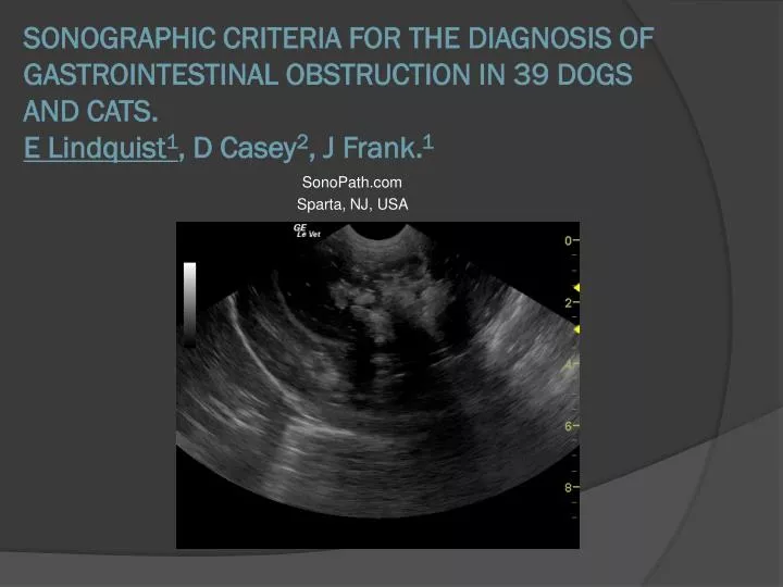
SonoPath.com Sparta, NJ, USA. SONOGRAPHIC CRITERIA FOR THE DIAGNOSIS OF GASTROINTESTINAL OBSTRUCTION IN 39 DOGS AND CATS. E Lindquist 1 , D Casey 2 , J Frank. 1. Purpose. Propose repeatable sonographic criteria for the diagnosis of gastrointestinal obstruction in dogs and cats.

E N D
SonoPath.com Sparta, NJ, USA SONOGRAPHIC CRITERIA FOR THE DIAGNOSIS OF GASTROINTESTINAL OBSTRUCTION IN 39 DOGS AND CATS.E Lindquist1, D Casey2, J Frank.1
Purpose Propose repeatable sonographic criteria for the diagnosis of gastrointestinal obstruction in dogs and cats.
Study DesignRetrospective analysis was performed on sonograms in 39 cases of dogs and cats that were found to have gastrointestinal obstruction upon surgical laparotomy.
Sonographic Parameters Analyzed • Proximal luminal gastrointestinal dilation • Proximal gastrointestinal hyperperistalsis • Presence of a foreign object/dysfunctional tissue at the end of the dilation • Type of acoustic shadow at obstruction site • Empty post obstruction intestinal lumen
Normal Parameters Identify Empty Colon and Small Intestine Normal Colonic Dirty Shadow
Complex Causes Of Obstruction Underlying Neoplasia Dysfunctional Bowel
Results • Proximal luminal gastrointestinal dilation was present in 39/39 cases. • Proximal gastrointestinal hyperperistalsis was present in 34/39 cases. The remaining 5 cases were determined to possess mechanical ileus owing to “exhausted bowel.” • An obstructive foreign object or tissue was discovered at surgery In 39/39 cases. • Acoustic shadowing was strongly present in 30 cases. Mild acoustic shadowing or “dirty shadow” similar to colonic content was present in 7 cases. Two cases did not present with an acoustic shadow on ultrasound. • Empty post obstruction intestinal lumen was definitively evident in 27/39 cases. Post obstruction fluid was present in 2 cases, and undecided in 10 cases.
Things To Consider • Sonographer experience • Feeding history • Systemic disease/hydration • Time elapsed between sonogram and surgery
Conclusions • From this study group of 39 cases, a consistent set of sonographic criteria for gastrointestinal obstruction can be utilized and combined with clinical signs and other testing in order to select medical versus surgical therapy for the patient presenting with gastrointestinal signs.
Additional Presentations To Be Considered • The “dirty shadow” of the colon was identified and followed to the ileocecal valve in order to differentiate it from dilated small intestine. • Obstructive presentations differed in character depending on sono-absorptive and fluid-absorptive characteristics: Corn cob vs. fabric vs. wood…… • 2 cases lacked an obstructive foreign object but were “obstructing” owing to dysfunctional bowel. • Periserosal fat inflammation and loss of serosal detail were found to be consistent with inflammation noted at surgery and was used as an “urgency factor.”