Download

1 / 74
740 likes | 778 Views
Leukemia. 浙江大学血液病研究所 医学院附属一院血液科 金洁. Definition. Leukemia is a clonal, malignant disease of hematopoietic tissue that is characterized by: (1) the proliferation of abnormal leukemic cells, principally in the marrow; (2) impaired production of normal blood cells.

E N D
Leukemia 浙江大学血液病研究所 医学院附属一院血液科 金洁
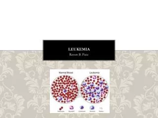
Definition Leukemia is a clonal, malignant disease of hematopoietic tissue that is characterized by: (1) the proliferation of abnormal leukemic cells, principally in the marrow; (2) impaired production of normal blood cells. (3)infiltration in tissue and organs Thus, the leukemic cell infiltration in marrow is accompanied, nearly invariably, by anemia and thrombocytopenia. The absolute neutrophil count may be low or normal, depending on the total white cell count. Also, Hepatomegaly, Splenomegaly and lymphopathy because of infiltration in tissue and organs
Incidence about 3-4 per 100,000; It is more common in males. About 1.81:1. AML about 1.6 per 100,000; ALL about 0.69 per 100,000; CML about 0.39 per 100,000; CLL about 0.05 per 100,000
Etiology Virus: HTLV-1 Chemical and drugs: Benzene, Alkylating agents and other cytotoxic drugs Radiation Inherited conditions: Identical sibling with Leukemia; Down syndrome; Fanconi anemia Double hits
Classification: Acute Leukemia: AML, ALL Chronic Leukemia: CML, CLL Special kind of Leukemia: HCL
Acute Leukemia MIC-M: Acute leukemia classification should now be based on Morphology, Immunophenotyping, Cytogenetic analysis and Molecular genetic analysis, namely MIC-M classification. Classification: FAB classification: ANLL(AML): M0-7 ALL: L1-3 MIC-M Classification: WHO: t(15; 17) Leukemia, t(8; 21) Leukemia
FAB classification M0: minimally differentiated AML M1: AML without maturation M2: AML with maturation M3: acute promyelocytic leukemia, APL M4: acute myelomonocytic leukemia, AMMoL M4Eo: AML with eosinophilia M5: acute monocytic leukemia M6: erythroleukemia, EL M7: acute megakaryoblastic leukemia
Bone Marrow of Acute myeloid leukemia (M1): The myeloid blast >90%, were round or oval, somewhat larger, abundant cytoplasm of opaque blue and non-particle. We can see some large vacuoles. Nucleus were oval or irregular in shape, sometime with large and clear nucleoli, and the nuclear staining particles were of small size.
Bone Marrow of Acute myeloid leukemia (M2): type 1 and type 2 blasts were >30%. Progranulocyte and its myeloid following were >10%. Auer body can sometimes be observed. There are three cells in the picture above have Auer body in their cytoplasm.
急性粒细胞白血病(M2b)骨髓象:此型据天津血液学会议建议暂定名,原称为亚急性粒细胞白血病。片中以体积增大,核浆发育不平衡的中幼粒细胞增生为主。这类细胞已发育为中幼粒阶段,而胞核发育落后于胞浆。急性粒细胞白血病(M2b)骨髓象:此型据天津血液学会议建议暂定名,原称为亚急性粒细胞白血病。片中以体积增大,核浆发育不平衡的中幼粒细胞增生为主。这类细胞已发育为中幼粒阶段,而胞核发育落后于胞浆。
Bone marrow Acute myeloid leukemia (M2b): This type was temporarily named according to the proposal of Tianjin Hematology meeting, formerly known as a sub-acute myeloid leukemia. Myelocytes of increased volume and of nuclear-cytoplasmic development imbalance were mainly observed. Such cells have been developed to promyelocytic stage, but the development of nucleus was later than cytoplasm.
(接上页)有的细胞核染质细致疏松,核仁明显,似仍处于原粒阶段。胞浆中颗粒减少,分布不匀,或出现空泡。部分早幼粒细胞也同样可出现核浆发育不平衡的现象。本例骨髓中红系细胞增生活跃,部分幼红细胞呈巨幼样变。(接上页)有的细胞核染质细致疏松,核仁明显,似仍处于原粒阶段。胞浆中颗粒减少,分布不匀,或出现空泡。部分早幼粒细胞也同样可出现核浆发育不平衡的现象。本例骨髓中红系细胞增生活跃,部分幼红细胞呈巨幼样变。
(Continue). Some nuclei are with small, loose particles and obvious nucleolus, as if they were still in the myeloblast stage. Plasma particles decreased and distributed unequally, sometimes vacuoles can be seen. Nuclear-cytoplasmic development imbalance was oberved in part of promyelocyte. Erythroid line actively proliferated in this case, and megaloblastoid erythropoiesis could be observed.
Acute promyelocytic leukemia (M3) Bone marrow: abnormal promyelocytes with abundant particles proliferate significantly, regular >90%. They’re irregular in size and shape, with a clear pseudo-foot protruding. Some are obvious two layers of cytoplasm, the plasm within maitain light red, covered with coarse, different shapes of Legionella Azure particles; Plasm was blue outside, often non-particles. Nucleus are of irregular shape, nuclear staining particles are crude, without nucleolus or 1-2 small nucleoli.
Acute promyelocytic leukemia (M3b, or fine particle type) myelogram: small particles in cytoplasm, still a small number of cells containing the typical coarse particles, and Auer body can be observed.
M4 bone marrow: blast cells (smaller size) increased. naive monocytes can be observed with larger size and cytoplasm of faded blue, nuclei are of irregular shape, some were folded or depression, nuclear staining particles are fine and loose, we can see one or two nucleolus.
Acute monocytic leukemia (M5a type) myelogram : The proliferation of monocytes (significantly more than 80%). Cells were round, with blue gray cytoplasm, pseudopod-like protuberances within the outer, no particles. Most were with round nuclear and fine nuclear staining particles, we can see one or two nucleolus.
Acute monocytic leukemia (M5b type) myelogram : promonocyte >20%, irregular in shape, with pseudo-foot, Nuclear staining particles are fine and loose, we can see nucleolus, but the cytoplasm shows the small purple particles, some have lots of particles more throughout the cytoplasm.
AML-M6 : significant erythroid proliferation, and more than 50%, and with morphological changes. Primitive myeloid blasts over 30% (NEC classification). Myeloblast and normoblast increased in the picture above, with megaloblastoid erythropoiesis.
M6 bone marrow: Super-giant red blood cells can be observed. Giant RBCs are about 30-35 microns in diameter, was stained of same colous with other red blood cells. The picture above showed a cell with multi-stained color uniformity and without central olistherozone.
Small promegakaryocyte and micromegakaryocyte which produces platelet.
FBA classification for ALL L1: small cell ≥ 12um L2: large cell > 12um L3: Burkit type
Acute lymphoblastic leukemia (L2) bone marrow: the large lymphocytic blasts with large amount of cytoplasm. Irregular-shaped nuclei with large and visible nucleolus.
Clinical features: Signs&symptoms (1) General: 1. pallor, fatigue, weakness, palpitation, dyspnea on exertion (they reflect the rise of anemia) 2. Easy bruising, petechiae, epistaxis, gingival bleeding ( they reflect thrombocytopenia) 3. Fever is present in many patients at the time of diagnosis ( absolute neutrophil counts under 500/ul) 4. enlargement of the liver, spleen or lymphnodes
Clinical features: Signs&symptoms (2) Special: Widespread bleeding caused by DIC (mainly in M3) Gum hypertrophy (mainly in M5, M4) Bone or joint pain Headache, confusion and dyspnea caused by hyperleukocytosis (hyperleukocytotic leukemia) Chloroma
Clinical features: Laboratory Findings(1) Blood routine hyperleukocytotic leukemia: circulating WBC> 100,000/μL hypoleukocytotic leukemia: circulating WBC<1,000/μL Hb Plt
Clinical features: Laboratory Findings(2) Bone marrow routine Leukemic blast≥20%(WHO), ≥30% (FAB) If blast cells is < 20%, there is t(15;17); t(8;21); t(16;16)/inv(16) Auer body A rod-shaped structure in the cytoplasm of immature myeloid cells, especially myeloblasts, in cases of acute myelocytic leukemia. Hiatus (gap) phenomenon裂孔现象 Chemical Staining: MPO; SB; NSE (NaF inhibit)
Clinical features: Laboratory Findings(3) Immunophenotyping Cytogenetic analysis t(15;17)(q22;q21) PML/RARa inv(16)(p13;q22) CBF-Myh11 t(16;16) (p13;q22) t(8;21)(q22;q22) AML/ETO t/del(11)(q23) MLL/ENL
Histochemical stain for AML-M5: nonspecific esterase (NSE) positive suppressed by NaF
Cytogenetic change in AML-M3: t(15;17)
Differential diagnosis Acute aplastic anemia idiopathic thrombocytopenic purpura infectious mononucleosis myelodysplastic syndrome
Treatment Combined chemotherapy induction chemotherapy: AML DA, HA,HAA ALL VDCP, VDLP M3 ATRA, Arsenic Trioxide ~~~~~~~~~~~~~~~~~~~~Why? Supportive treatment blood transfusion, G-CSF/GM-CSF, antibiotics Consolidation chemotherapy Stem cell transplantation
Prognosis Cytogenetics Age WBC count during disease onset Immunophenotyping
Hierarchical Cytogenetic Prognostic Classification • Analysis of 1,612 patients derived from MRC AML 10 (median age 35 years; range, 0–55 years) based on CR, RR, and OS • FAVORABLE • t(8;21), t(15;17), inv(16), either alone or in conjunction with other changes • INTERMEDIATE • Normal, +8, +21, +22, del(7q), del(9q), abnormal 11q23, or other structural/numerical abnormalities • Lack of additional favourable or adverse abnormalities • ADVERSE • -5, del(5q), -7, t( 3;3), complex (>3 unrelated changes) • alone or in conjunction with intermediate-risk or other • adverse-risk abnormalities Grimwade D, et al. Blood. 1998;92:2322-2333.
MRC AML 10: Survival From Start of Course 2 by Cytogenetic Group 75% 42% 18% P<0.0001
Risk Group Definition RiskgroupMRC US Intergroup Good M3, t(15;17) t(15;17) with other: inv(16) t(8;21) inv(16) t(8;21) without del (9q) or complex Standard not good or poor +8, -Y, +6, del(12p) normal Poor -5/del(5q); del(7q) -5/del(5q), -7/del(7q), inv(3q), t(3;3), complex 11q, 20q, 21q, 17p, del(9q), t(6;9), t(9;22) complex Slovak ML, et al. Blood. 2000;96:4075-4083.
Chronic Leukemia Classification: CML, CLL, HCL (hairy cell leukemia) Age and gender of onset: CML: all age, commonly middle age male slightly more than female CLL: mainly elder male slightly more than female
chronic myelogenous leukemia Incidence: 0.39-0.99 per 100,000 Stage: Chronic phase CP; Accelerated phase; Blastic phase(Blastic crisis) BP/BC
Clinical manifestations fatigue, night sweats, low-grade fever syndrome caused by hyperleukocytosis splenomegaly