Download
1 / 68
740 likes | 808 Views
PHYSIOLOGY OF THE RENAL SYSTEM. Fredirick L Mashili, MD, PhD. DEPARTMENT OF PHYSIOLOGY, MUHIMBILI UNIVERSITY OF HEALTH AND ALLIED SCIENCES (MUHAS). CONTENTS. Kidney functions Structure of the kidney Vascularization of the kidney Juxtaglomerular apparatus Glomerular filtration

E N D
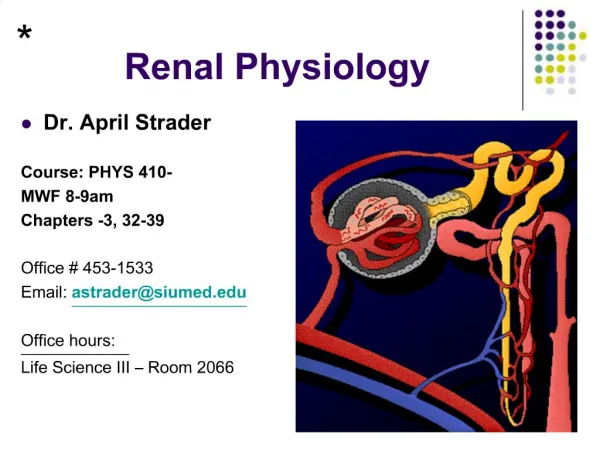
PHYSIOLOGY OF THE RENAL SYSTEM Fredirick L Mashili, MD, PhD. DEPARTMENT OF PHYSIOLOGY, MUHIMBILI UNIVERSITY OF HEALTH AND ALLIED SCIENCES (MUHAS)
CONTENTS • Kidney functions • Structure of the kidney • Vascularization of the kidney • Juxtaglomerular apparatus • Glomerular filtration • Tubular reabsorption • Tubular secretion • Ureter physiology • Urinary bladder • Micturition • References
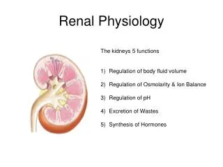
KIDNEY FUNCTIONS 1. HOMEOSTASIS • maintain the blood volume and the normal composition of body fluid compartments • excrete waste products ( urea, creatinine, uric acid, NH₃(ammonia), which are toxic for the organism • regulate the blood concentration of ions ( Na+, K +,Cl, Ca++, sulphate, phosphate, bicarbonate) • maintain the pH by secreting the H + • maintain the osmolarity and water volume via the capacity to adjust the water reabsorption • regulate arterial blood pressure • gluconeogenesis
KIDNEY FUNCTIONS 2. ENDOCRINE ROLE • synthesis of erythropoetin - sensory cells at the proximal convoluted tubules (PCT), which respond to changes in the partial pressure of oxygen (pO2). • role in metabolism of vitamin D and Calcium - active vitamin D needed to reabsorb Ca²+ in small intestine - to activate vitamin D: an additional hydroxyl group is added => 1.25 dihydroxycolecalciferol - Vitamin D pathway: 1. 7-dehydrocholesterol under the action of UV rays becomes colecalcipherol or vit. D3 ( in skin) 2. Vit. D3 in liver becomes 25 OH D3 and then in kidneys 1,25 (OH)2 D3 or calcitriol→ increase Ca absorption in the intestin
KIDNEY FUNCTIONS 2. ENDOCRINE ROLE RENIN- ANGIOTENSIN- ALDOSTERON SYSTEM (RAAS) • juxtaglomerular cells (in the wall of the afferent arteriole) synthesize the enzyme RENIN, a glycoprotein with 42000 D , that catalyses the transformation of angiotensinogen (from liver) into angiotensin I. • Angiotensin I is transformed into Angiotensin II ( reaction catalyses by angiotensin converting enzyme – in the lungs) • Angiotensin II causes vasoconstriction (especially in the skin, abdominal organs, kidney (acts on efferent arterioles); less in brain, muscles, heart • Angiotensin II stimulates ALDOSTERONE secretion (in adrenal gland) • Renin is released in case of: renal ischemia (decrease of blood supply to the kidney), decreased blood volume ( due to bleeding, dehydration), hypotension (low blood pressure (BP), cardiac failure
RENIN ANGIOTENSIN SYSTEM (after R.Rhoades & G.Tanner, Medical Physiology, 2003)
KIDNEY FUNCTIONS 2. ENDOCRINE ROLE • Release prostaglandins E₂(PgE₂), Pg F2 alpha and Pg I (Prostacyclin)- they act more in a paracrine manner - Pg E₂in hypertensive people : - decrease the blood pressure - increase : - renal blood flow and diuresis (volume of excreted urine/day) - natriuresis (amount of Na excreted via urine/day) - Pg F2 alpha => vasoconstriction
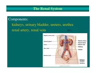
STRUCTURE OF THE URINARY SYSTEM The renal apparatus: 1) kidneys (produce urine) 2) urinary excretory pathways • ureters • urinary bladder (it accumulates and stores urine between 2 micturitions) • urethra
STRUCTURE OF THE KIDNEY • NEPHRON is the morphological and functional unit of the kidney - there are 1-1.2 millions of nephrons per kidney - it is made up of : 1) renal corpuscle and 2) tubule 1) Renal corpuscle or Malpighian corpuscle/body • Diameter of 200 μm • It is placed in the cortex of the kidney • It consists of : a) Capillary tuft (aprox. 50 capillaries) or glomerulus b) Bowman’s capsule • Role - at its level the process of plasma filtration (glomerular filtration) takes place => primary urine
STRUCTURE OF THE KIDNEY 2) Tubule - 45 - 65 mm For reabsorption and secretion processes • a) Proximal convoluted tubule (PCT) o length: 14-12 mm; diameter: 55 μm o one layer of columnar cells on a basal membrane o cells with brush border at the apical pole (towards the lumen) with many microvilli (for reabsorption- increased surface) o cells have invaginations at the basolateral pole (with a striated aspect and many mitochondria)
STRUCTURE OF THE KIDNEY b) Loop of Henle- like a hairpin • it has 2 limbs: ascending and descending, each with a thin and a thick segment • 15-20% of glomeruli have long loops of Henle going deep into medulla - 26mm • Cells: cuboidal in the thick limb and squamous in the thin limb • Macula densa: at the final portion of ascending limb the structure is modified (with bigger and fewer cells rich in mitochondria) - Cells with osmo- /chemoreceptors sensitive to Na and Cl concentrations in the urine • Juxtaglomerular apparatus: macula densa and juxtaGLOMERULARcells • When the conc. of Na or Cl in the macula densa decreases => takes place the release of renin from juxtaglomerular cells
STRUCTURE OF THE KIDNEY c) Distal convoluted tubule (DCT) o length: 5-8 mm; diameter: 30-40 μm o only a few microvilli, but without a brush border o several distal tubule together become 1 collecting duct (CD), which crosses the cortex and medulla, opens into the renal pelvis and continues into the ureters d) Collecting duct (CD) o across the cortex and the medulla of the kidney o concentrates urine o reabsorption of H2O under the influence of ADH o collection of urine from 3000-5000 nephrons/CD
STRUCTURE OF THE KIDNEY(after A.Despopoulos & S.Silbernagl , Color Atlas of Physiology, 2003)
VASCULARIZATION OF THE KIDNEY • By the renal artery (directly from the abdominal aorta) -> interlobar arteries (terminal arteries, without anastamosis; in case of an obstruction => necrosis of that territory results) -> arcuate arteries -> interlobular arteries -> afferent arterioles (enter the glomerolus) -> capillary tuft (50) -> efferent arterioles-> peritubularcapillaries (loop around the tubules) -> interlobular veins -> arcuate veins -> interlobar veins -> renal vein -> inferior vena cava • 2 networks of capillaries from the efferent arterioles 1) peritubular capillaries (collected by interlobular veins) 2) vasa recta (around the tubules ofthejuxtamedullary nephrons) • R-I-A-I-A-C-E-P-I-A-I-R • R --- ACEP --- R
JUXTAGLOMERULAR APPARATUS Juxtaglomerular apparatus is located in the hillum of every glomerulus • It is made up of juxtaGLOMERULAR cells and the macula densa • Modified muscular layer in the structure of afferent and efferent arterioles: - increased number of smooth muscle cells of afferent arterioles (Af. A) as long as the arteriole comes closer to the glomerulus - cells become thicker (with less actin/myosin filaments, but with many granules containing RENIN) - cells of the juxtaglomerular apparatus act as baroreceptors sensing changes in BP (cells are stimulated by distension of the afferent arteriole; if not distended - release of RENIN) • Low BP (wall of Af. A. is not distended) => Renin secretion => Angiotensin II => increase reabsorption of Na and water => increase BP
JUXTAGLOMERULAR APPARATUS (after Vander et al., Human Physiology: The mechanisms of body function, 2001)
RENAL CIRCULATION AND ITS REGULATION • Renal circulation has the capacity of autoregulation • It is an intrinsec property of the kidney => it is observed even on isolated, denervated kidney • It is necessary for maintaining a constant GFR and excretion of water and waste products • BP CHANGE OF = 80 –180 mmHg => a constant GFR abd RBF ( renal blood flow) are maintained • It prevents the high variation of water and solutes excretion together with the increase of BP • Autoregulation is explained by two mechanisms: 1) tubuloglomerular feedback mechanism 2) myogenic mechanism
AUTOREGULATION OF THE RENAL CIRCULATION 1) Tubuloglomerular feedback mechanism • The increase in BP => increase in GFR => increased NaCl delivery to the macula densa => increased NaCl reabsorption by macula densa cells => constriction of afferent arteriole results • Vasoconstriction can be mediated by : Nitric oxide (NO) , adenosine, ATP • => Renal blood flow (RBF), GFR are lowered to a more normal value. • The tubuloglomerular feedback mechanism = a negative-feedback system that stabilizes RBF and GFR. • Tubuloglomerular feedback mechanism controls the amount of Napresented to distal nephron segments, because these segments have a limited capacity to reabsorb Na. • Renal autoregulation minimizes the impact of changes in arterial blood pressure on Naexcretion. • Decreased macula densa sodium chloride causes dilation of afferent arterioles and increased Renin release • Without renal autoregulation, increases in arterial blood pressure => increases in GFR and losses of NaCl and water from the ECF.
THE TUBULOGLOMERULAR FEEDBACK MECHANISM(after R.Rhoades & G.Tanner, Medical Physiology, 2003)
AUTOREGULATION OF THE RENAL CIRCULATION 2) Myogenic mechanism • The increase in BP=> stretches blood vessel wall => opening stretch-activatedcation channels in smooth muscle cells=> membrane depolarization=> opening voltage-dependentCalciumchannels=>intracellular [Ca] rises=>smooth muscle contraction =>vessel lumen diameter decreases=>vascular resistance increases • Decreased BPinduces the opposite changes
GLOMERULAR FILTRATION • First step in urine formation (reabsorption and secretion follow) • 25% of the plasmatic renal flow are filtered in the Bowman’s capsule (primary urine) • in resting condition, the tow kidneys receive 1.2-1.3 L/min of blood (=25% of the cardiac output); of this 25% is filtered (only H2O, micromolecules, small proteins, no blood cells or substances bound by plasma proteins) • Primary urine: 180 L/day, with a similar composition as plasma • Final urine: 1.0 – 1.5 L/day, with a composition modified by reabsorption and secretion
GLOMERULAR FILTRATION MEMBRANE • Anatomical support for glomerular filtration. Structure (3 layers): • a) Endothelial cells of capillaries - the glomerular capillaries are fenestrated, with holes of 40-100nm in diameter. The endothelial cell surface and the holes are covered with a glycoprotein coat (glycocalyx), about 12 nm thick and with a negative electrical charge • b) Basement membranehas 3 layers : 1. Internal lamina rara 2. Lamina densa (more dense middle layer) 3. External lamina rara It is made up of proteoglycans and collagen fibers, with large spaces through which water and micromolecules can pass. Thickness: 310-340nm • c) The epithelial cells of Bowman’s capsuleare named podocytes with many footlike extensions. Podocytes are fixed on the external lamina rara by pedicels. Between the pedicels there is a thin membrane of 4-6 nm in thickness, named slit membrane. - Pores of the slit membrane: 20-30 nm - The surface of the podocytes and slit membrane is covered with glycocalyx. Due to the negative electrical charge, proteins are repelled and their passage into the urine is prevented. This process can be disturbed in many renal diseases (albumin can pass into the urine => albuminuria)
GLOMERULUS AND BOWMAN`S CAPSULE( after A.Despopoulos & S.Silbernagl, Color Atlas of Physiology, 2003)
FACTORS INFLUENCING GLOMERULAR FILTRATION (GF) 1) Permeability of the membrane • Some substances have been used for studies of glomerular filtration membrane permeability : - anionic ferritin - 6.1 nm in diameter, negative electrical charge; it cannot pass through the lamina densa of the basement membrane - cationic ferritin passes easier than anionic ferritin, but it is stopped by slit membrane and podocytes - colloidal gold - dextrans (colloid substances used in perfusions to recover BV) • Molecules bigger than albumin (69000 D) are stopped by slit membrane - Plasma albumin: 0.2% passes into urine - Haemoglobin: 5% passes into urine (less negatively charged than albumin) • The smaller and more positively charged the particles are, the easier they can pass through the filtration membrane • Substances with MW < 10000 D can be filtered • Substances bound by plasma proteins ( Ca 2+, free fatty acids) are not filtered • Glomerulonephritis - proteins can pass due to altered permeability (modified glycocalyx amount/structure) => albuminuria
FACTORS INFLUENCING GLOMERULAR FILTRATION 2) Surface area of filtration • for the two kidneys is 1.2-1.5 m² • active surface area depends on the number of working nephrons (in humans all nephrons work all the time) • decreased filtration surface/rate: - when mesangial cells among the capillaries of the glomerolus contract (by Angiotensin II and ThromboxanE A₂) - when podocytes are relaxed /flattened they cover more of filtration surface - in many renal diseases reduce the filtration surface (nephrons are destroyed)
FACTORS INFLUENCING GLOMERULAR FILTRATION 3) Effective filtration pressure • The volume of filtered plasma depends on the difference between the hydrostatic pressure and the colloid osmotic pressure of the blood and Bowman’s capsule • Pressure of filtration = hydrostatic pressure of the blood (HPB) – hydrostatic pressure of Bowman’s capsule (HPBC) – colloidosmotic pressure of blood (CPB) Filtration pressure = 45mmHg (HPB)- 10mmHg (HPBC)- 25mmHg (CPB) = 10mmHg Hydrostatic pressuredecreases with only 1-2 mmHg throught the length of capilllaries, but colloid osmotic pressure increases significantly • Variations - Glomerular filtration=>variation if BP changes between 80-180mmHg - Shock/collapse => glomerular filtration decreases (vasoconstriction at the afferent and efferent arterioles). GF stops when BP < 40-50mmHg - Vasoconstriction of the afferent arterioles => decreased glomerular filtration (by CATECHOLAMINES, etc.); at efferent arterioles => increased gomerular filtration (by ANGIOTENSIN II, etc.) - Increased hydrostatic pressure in Bowman’s capsule => impeded filtration (stones, tumors, edema in the renal parenchyma) - Colloidosmotic pressure Decreased => increased filtration (hypoproteinemia) Increased => decreased filtration (hyperproteinemia)
REGULATION OF GFR and RBF • GFR is influenced by : 1) sympathetic nervous sytem 2) hormones 3) autacoids 1) Sympathetic nervous system (SNS) Afferent and less efferent arterioles receive sympathetic fibers • Strong stimulation of SNS => constriction of afferent A. => decreases RBF => decreases GFR • Moderate stim. of SNS => little influence of GFR • Its role is more important in : bleeding, shock, ischemia and less in normal conditions
REGULATION OF GFR and RBF 2) Hormones • Norepinephrine, Epinephrineconstrict renal blood vessels (afferent and efferent A.) and decrease GFR; are released from adrenal medulla. Normally they have little influence on renal blood flow, except some acute conditions (bleeding) • Angiotensin II constricts afferent arteriole; its formation increases in circumstances associatedwith decreased arterial pressure or volumedepletion, which tend to decrease GFR. • The increased level of angiotensin II=>constriction of efferent arterioles=> increases GFR => maintains normal excretion of metabolic waste products ( urea and creatinine) that depends on GFR for their excretion Angiotensin II, by stimulating the secretion of Aldosteron=> increases tubular reabsorption of sodium and water => restores blood volume and blood pressure
REGULATION OF GFR and RBF 3) Autacoids • Endotelin - produces vasoconstriction of renal blood vessels - increases intoxemiaof pregnancy, acute renal failure, and chronic uremia=> decreases GFR • Endothelial-Derived Nitric Oxide (NO) - decreases renal vascular resistance and increases GFR - it is important for maintainingvasodilation of the kidneys - administration of drugs that inhibit this normalformation of NO => increases renal vascularresistance and decreases GFR and urinary sodiumexcretion=>high BP • Prostaglandins (PGE2 and PGI2) and Bradykinin => Increase GFR -Prostaglandins may help prevent excessive reductionsin GFR and renal blood flow under stresfull conditions: volume depletion or after surgery - the administration of nonsteroidal anti-inflammatoryagents (Aspirin), that inhibit prostaglandin synthesis=> reduction in GFR
TUBULAR REABSORPTION • During the passage of filtrate through the renal tubule => its composition is changed • Substances move from the tubule to the peritubular capillaries = tubular reabsorption and • from peritubular capillaries to the tubular lumen = tubular secretion • Tubular reabsorption by : - passive transport - active transport
TUBULAR REABSORPTION OF GLUCOSE • It takes place at the level of proximal tubules (PCT) • 98% of filtered glucose is reabsorbed at PCT (cca.100g/day) GFR: 125ML/MIN, normal glycemia: 80-120mg% => Glucose filtered/min: 125mg/min (filtered load) • The reabsorbed amount of glucose depends on the amount of filtered glucose and the capacity to transport glucose • Glucose transport capacity has a maximum = 375mg/min in males and 300mg/min in females • If maximum limit is exceeded => glucosuria (glucose excreted into the urine) • Glucose threshold = glucose plasma concentration at which the maximum capacity of reabsorption is exceeded and glucosuria occurs ; It is 180mg% for venous blood and 200mg% for arterial blood Not all nephrons have the same filtration and reabsorption capacity • Reabsorption - by symport with Na - at the apical membrane of tubular cells (secondary active transport , mediated by SGLT 1). Glucose diffuses to the interstitial space (by GLT 2) and Na is pumped to the interstitial space (via Na-K-ATPase). Galactose and fructose are competitors for the glucose-symport • Renal diabetes - glucose reabsorption is reduced/ impeded ( normoglycemia with glucosuria) • Diabetes mellitus -with an overwhelmed glucose reabsorption capacity (hyperglycemia with glucosuria)
REABSORPTION OF GLUCOSE AND OTHER ORGANIC SUBSTANCES (after A.Despopoulos & S.Silbernagl, Color Atlas of Physiology, 2003)
TUBULAR REABSORPTION OF AMINO ACIDS AND PROTEINS • Amino acids (Aa) are reabsorbed at the level of PCT. Daily 70 g of Aa are filtered . • It is similar to glucose reabsorption (Na coupled secondary active transport) • Almost complete reabsorption (maximum 1-2% excreted into the urine) • There are described several transport systems/ carriers: 1. transport of neutral amino acids (diaminic Aa) 2. transport of proline and hydroxyproline 3. transport of β-amino acids 4. transport of diaminic Aa (arginin, lysine) and dicarboxylic Aa (aspartic acid, glutamic acid) • Defects in reabs. of some Aa => cystinuria (L-cystine, L-arginineand L-lysine are hyperexcreted) => urinary calculus • Proteins- especially albumin, but also lyzozyme, alpha 1-microglobulin, beta 2-microglobulin are filtered - reabsorption - by receptor mediated endocytosis. Proteins are digested by lysosomes inside the cells of the renal proximal tubule, split into aminoacids, which are reabsorpted - this typeof reabsorption is nearly saturated at normalfiltered loads of proteins=>an elevated plasmaprotein conc. or increased protein sievingcoefficient=>proteinuria
REABSORPTION OF OLIGOPEPTIDES AND PROTEINS (after A.Despopoulos & S.Silbernagl, Color Atlas of Physiology, 2003)
TUBULAR REABSORPTION OF UREA AND URIC ACID UREA - daily formed: 25-30g (waste product of protein metabolism) • 30-90% reabsorbed (according to diuresis and density of urine) • At PCT: 60-65% of water reabsorbed (isoosmotic reabsorption) => urea concentration gradient is obtained • daily filtered: 54g of urea => daily reabsorbed: 30g • if urinary flow > 2mL/min => 60-70% of urea is reabsorbed • Urea reabsorption occurs also at the DCT and CD under ADH action URIC ACID • Plasma conc. = 3 –7 mg% • It is both reabs. and secreted in PCT • waste product of nucleoprotein catabolism • daily excreted - 10% of filtrated uric acid = 1g/day • alkaline pH => uric acid from urine found as salts (urate - Na urate, K urate) • acidic pH => uric acid found as acid (uric acid) => stones formed • pH measurement if there is a kidney stone suspicion
REABSORPTION OF UREA (after R.Rhoades & G.Tanner, Medical Physiology, 2003)
TUBULAR REABSORPTION OF SODIUM • 1kg NaCl is filtered daily • all the segments of the nephron participate in Na reabsorption (except the descending limb of the loop of Henle) • Na reabsorption by diffusion ( along an electrochemical gradient), by symport with glucose and counter transport with H • from cells Na is pumped into the interstitial space (via Na-K-ATPase) • if Na-K-ATPase is blocked by Oubaine => Na reabsorption in the PCT is reduced by 50% • Na ions accumulate in the interstitial space => hyperosmotic environment which attracts H2O to the interstitial space by osmosis (cells at the PCT are permeable to H2O without hormonal intervention -ADH) Cl anions follow Na movement • 65% of filtered Na is reabsorbed at the PCT (along with diffusion of H₂O from the interstitial space to the vessel) • 25% of filtered Na is reabsorbed at the ascending limb of the loop of Henle • 8-10% of filtered Na is reabsorbed at the DCT and CD
ION TRANSPORT IN THE THICK ASCENDING LIMB (after R.Rhoades & G.Tanner, Medical Physiology, 2003)
CONTROL OF SODIUM REABSORPTION • Na - the main cation of extracellular fluid • It produces 90% of the osmotic pressure of the extracellular fluid compartment • Na concentration it is precisely regulated => it gives extracellular fluid volume • Na intake is variable - 8-10g/day • Renal excretion of Na is regulated by : - ALDOSTERONE - mineralocorticoid hormone produced by thesuprarenal glands - increases Na reabsorption in exchange with K and H, which are secreted - CORTISOL increases Na reabsorption - ATRIAL NATRIURETIC PEPTIDE (ANP) - secreted by atrial myocytes; increases natriuresis (excretion of Na ) and GFR • amount of Na reabsorbed varies proportionately with renal flow • decreased Na or Cl concentrations at DCT stimulate macula densa cells followed by a release of RENIN (by the juxtaglomerular apparatus), => stimulates the formation of ANGIOTENSIN II => stimulates the ALDOSTERONE formation => increases the reabsorption of Na
TUBULAR REABSORPTION OF CALCIUM • Ca is not completely filtered because a fraction of plasma Ca is bound to proteins in blood (40%- nonfiltrable) • Daily are filtrated 10000mg • 99.5% of filtered Ca is reabsorbed • 65% reabs. in PCT, 25-30% in LH, 4-9% in DT and CT • Parathyroid gland produces Parathormone (PTH) • PTH increases Ca reabsorption in DCT and CD • Acidosis (proteins bind less Ca) => increased amount of Ca filtered => calciuria
TUBULAR REABSORBTION OF CHLORINE • Cl is the main anion of the extracellular fluid • Reabsorption depends to a great extend on Na reabsorption ( Cl follows Na movement) • At the thick ascending limb of the loop of Henle - active Cl transport from the lumen to the cytoplasm along with Na and K via a carrier protein (Na-K-2Cl- transporter) • Inhibition by diuretics e.g. FUROSEMID (inhibits Na reabsorption)
TUBULAR REABSORPTION OF BICARBONATE • Not reabsorbed as HCO₃(bicarbonate), because in the presence of H: HCO₃+ H => H2CO₃ and H₂CO₃ => H₂O + CO₂ • CO₂ diffuses from the blood into tubular cells • Acidosis: entire filtered HCO₃is reabsorbed (under acid-base-balance only 99%) • Alkalosis: more HCO₃excreted, less reabsorbed
TUBULAR REABSORPTION OF BICARBONATE (after R.Rhoades & G.Tanner, Medical Physiology, 2003)
TUBULAR SECRETION OF HYDROGEN • Role - to maintain the normal blood pH • Acids react first with a buffer system => neutralization (mainly by NaHCO₃) • E.g. by RBCs: acid + NaHCO₃ => sodium salt of acid + H₂CO₃ • H₂CO₃ => CO₂ and H₂O • CO₂is removed by respiration and sodium salt removed through the urine • With every mole of filtered urine also 1 mole of NaHCO₃ is lost • Kidneys secrete H ions until the urinary pH of 4.5 is achieved • Secretion of H in PCT, DCT and CD
TUBULAR SECRETION OF HYDROGEN • PCT secretion via secondary active transport (antiport/countertransport) with Na • 1 H secreted and 1 Na + 1 HCO₃reabsorbed At apical membrane of tubular cells • DCT • H secreted by active transport (without Na- H-ATPase)- H pumps • H pumps stimulated by ALDOSTERONE (hormone produced by the suprarenal glands; role in reabsorption of Na + H₂O and secretion of H + K) • Acidosis => pumping/secretion of H • Alkalosis => pumping/secretion of K
TUBULAR SECRETION OF HYDROGEN • COLLECTING TUBULE • Secretion via 2 types of cells (intercalated and principal cells): 1)Intercalated cells- • o similar to the parietal cells of the stomach • o rich in carbonic anhydrase (catalyses CO₂ + H₂O => H₂CO₃) • o luminal membrane with ATPase for H secretion into urine 2) Principal cells production of H in the cells: - by dissociation of H₂O - using CO₂ as a source of H (CO₂ (by cell metabolism) -> blood-> tubular cells where CO₂ + H₂O => H₂CO₃ => H + HCO₃(H secreted in the urine and HCO₃is reabsorbed in the blood)) • H ions in urine react with buffer systems such as HCO₃, phosphates or NH₃to form H₂CO₃, monobasic phosphates or NH₄ • Most H secreted in the PCT reacts with HCO₃ and flows as H₂CO₃ trough urine H₂CO₃ => H₂O and CO₂ (which diffuses back to cells, reacts with H₂O => H₂CO₃