Download
1 / 21
230 likes | 730 Views
Renal physiology I. Lecture 36 Monday, April 2, 2007 Refs. Ross Chapter 20, Wheater’s Chapter 16, Medical Physiology Chapters 32 and 33 and Ganong Chapter 38. General steps in the formation of urine. Filtration of plasma produces ultrafiltrate

E N D
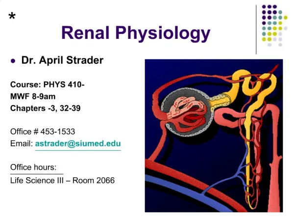
Renal physiology I Lecture 36 Monday, April 2, 2007 Refs. Ross Chapter 20, Wheater’s Chapter 16, Medical Physiology Chapters 32 and 33 and Ganong Chapter 38
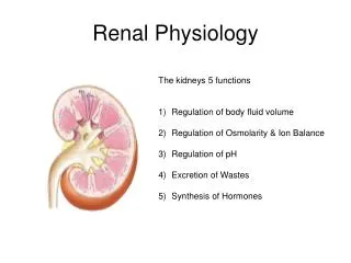
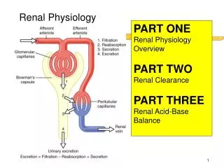
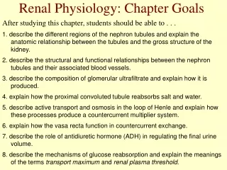
General steps in the formation of urine • Filtration of plasma produces ultrafiltrate • Total blood volume circulates through kidney ~300 times per day and cortex receives 90% • Selective reabsorption of electrolytes and nutrients from ultrafiltrate (water follows). • Secretion of some compounds into urine. • Secretion of H+ maintains acid-base balance. • Fluid balance (loss or conservation of water) is determined by permeability of the collecting duct.
Step 1: glomerular filtration • Filtration is a physical process. • As blood flows through the glomerulus, water and small solutes filter through the endothelium, basement membrane, and slit pores into the urinary space of Bowman’s capsule. • Whether a molecule enters the filtrate depends on its size, charge, and configuration. • Generally, molecules with weights ≤ 65,000 and a diameter < 4 nm pass through the filter. • The endothelium, basement membrane, and podocyte all have a negative charge. Neutral molecules up to 8 nm pass through. A negatively charged molecule close to the size limit will be held back.
Protein in urine • Normally, cells and plasma proteins remain in the blood. • Albumin has molecular weight of 68,000, a diameter of 7 nm, and is negatively charged. • Hemoglobin with a molecular wt. of 65,000 passes through filter. • Protein in the urine is normally < 100 mg/dL • mostly due to shed tubular cells • Thick ALH cells secrete a mucoprotein 30-50 mg/day • In nephritis, the negative charges in the glomerular wall are dissipated, and albumen can pass through resulting in albuminuria. • Elevated protein in urine = proteinuria
Pressures and filtration • Filtration depends on hydrostatic and osmotic pressure gradients. • Glomerular hydrostatic P = 60 mm Hg • Glomerular osmotic P = 32 mm Hg • Capsular hydrostatic P = 18 mm Hg • Capsular osmotic P = 0 • Effective filtration pressure • The difference between glomerular and capsular pressures is (60-32)-(18+0)=10 mm Hg • The main factor determining the pressure gradient is the hydrostatic pressure of glomerular blood. • Hydrostatic pressure in glomerulus depends on: • Systemic blood pressure • Resistance through the glomerular capillaries
Quantitative differences in filtration • Hydrostatic pressure in glomerular capillaries is higher than systemic capillaries • Because the afferent arterioles are short. • Because the efferent arteriole has a smaller diameter than the afferent arteriole. • Filtration also depends on the size of the capillary bed in the glomerulus. • Contraction of mesangial cells can reduce the area available for filtration. • Angiotensin II is an important regulator of mesangial cell contraction. • Many mediators affect mesangial cells.
Glomerular filtration rate (GFR) • The rate of formation of filtrate (ml/min). • GFR for an average-sized normal man is 125 ml/min. • GFR can be determined by measuring the plasma and urine levels of a substance that is freely filtered, not reabsorbed, and not secreted. • Inulin (a polymer of fructose with a mol.wt of 5200) is used. • The amount excreted in the urine over a period of time is equal to the milliliters of plasma that contained that amount. Factors affecting GFR are listed in the following table
Juxtaglomerular apparatus affects GFR • Tubuloglomerular feedback • Macula densa serves as chemoreceptor. • Increased NaCl in DCT causes constriction of afferent arteriole and thus decreased GFR. • Alteration of systemic blood pressure via renin-angiotensin system • Afferent arteriole acts as baroreceptor. • Decreased stretch in wall of arteriole causes greater release of renin into blood. • Renin converts angiotensinogen to angiotensin I which is converted to angiotensin II by endothelial ACE. • Angiotensin II increases systemic arterial pressure. • Increased pressure in afferent arteriole increases GFR.
Clearance • Tubular cells may add more of a substance to the filtrate or reabsorb a substance. • Tx is net amount of x transferred. • Clearance equals GFR if there is no secretion or reabsorption. • Clearance exceeds GFR is there is net tubular secretion. • Clearance is less than GFR is there is net tubular reabsorption.
Clearance formula • Cx = Ux * V/Px • Ux is concentration of X in urine. • V is the volume of urine formed per unit time. • Px is the concentration of X in plasma. • Cx is the volume of blood that would be totally cleared of the substance X per unit time. Actually, a larger volume of blood is partially cleared. • Units are usually ml/min. • Also Cx can be thought of as the virtual volume of plasma (per unit time) that would be needed to supply the amount of solute to the urine. • Urinary excretion rate = Ux * V • units are mg or moles/min
Step 2 of urine formation: reabsorption • About 99% of the filtrate is reabsorbed.Table 38-5 • Much of the reabsorption takes place in the proximal convoluted tubule. • Cotransporters move Na and glucose or Na and amino acids into the tubular cells. Fig 38-11 • Main cotransporter for glucose takes 1 Na and 1 glucose (in small intestine it is 2 Na per glucose) • Secondary active transport- Na-K ATPase pumps Na out through basolateral membrane. • The proximal convoluted tubule is about 15 mm long. Glucose and amino acids are normally completely reabsorbed by its end. See Fig 38-9
Reabsorption of various solutes in the proximal tubule. TF/P, tubular fluid:plasma concentration ratioGanong 38-9
PCT reabsorption of glucose • Normally all filtered glucose is reabsorbed in the first part of the PCT. • If blood glucose is excessive, it can overwhelm the transporters and glucose will appear in the urine. • Threshold is about 200 mg/dl.
Top: Relation between the plasma level (P) and excretion (UV.) of glucose and inulin. Bottom: Relation between plasma glucose level (PG) and amount of glucose reabsorbed (TG).Ganong 38-10