Download
1 / 73
740 likes | 930 Views
Glomerulopathy and Interistitial nephritis with polycystic dis. By Dr Rasol M Hasan. Pathogenesis of glomerular injury. Antibody mediated injury In situ immune complex deposition Fixed intrinsic tissue antigens

E N D
Glomerulopathy and Interistitial nephritis with polycystic dis. By DrRasol M Hasan
Pathogenesis of glomerular injury • Antibody mediated injury • In situ immune complex deposition Fixed intrinsic tissue antigens NC1 domain of collagen type4 antigen [anti GBM-nephritis] Heymann antigen [membranous nephropathy] Mesangial antigens Circulating immune complex deposition Endogenous antigen[DNA,Nuclearproteins,immunoglobulins,IgA] Exogenous antigen [infectious agents,drugs] Cytotoxic antibodies Cell mediated immune injury Activation of alternative complement pathway
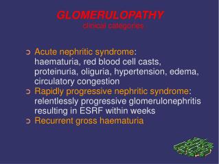
Clinical manifestation of glomerular injury • Asymptomatic • Macroscopic hematuria • Nephrotic Syndrome Nephritic syndrome • Rapidly Progressive glomerular nephritis • Chronic Nephritic Syndrome
Glomerular diseases with primary haematuria • IgA Nephropathy (Berger’s Disease] • Most common primary glomerular disease. • Mostly adolescents and young adults • gross hematuriaoccurring coincidentally with or immediately following (24-48 hours), a viral upper respiratory infection, flu-like illness, gastrointestinal syndrome • episodes of gross hematuria, • microscopic hematuria. • Focal and segmental glomerularmesangial proliferation, with IgA deposits. • Increased serum IgA. Normal C3 complement. • Prognosis – Generally benign • 20% progress to renal insufficiency in 10 years. • recurs after renal transplantation.
Membrano proliferative glomerulo nephritis (MPGN) Mesangiocapillary • 5-30 years • Immune complex disease • Associated conditions: Chronic infections (especially hepatitis C), cancer, heroin abuse, SLE, etc • Usually nephrotic syndrome, less often acute nephritic syndrome. Recent history of URI in many patients. Hypertension and/or renal insufficiency may occur. • Decreased serum complement levels. Hepatitis C serology should be obtained • Glomerularhypercellularity with capillary basement membrane thickening and splitting[TRAM-TRACKING]. Subendothelial deposits of C3 complement and sometimes IgG • . Prognosis Progressive deterioration of renal function; • Many patients develop end-stage renal insufficiency within 10 years.
Glomerular disease presenting as RPGN • Goodpasture’s syndrome • Vasculitis Wegner’s granulomatosis Microscopic polyangitis (MPA) Pauci immune crescentric glomerulonephritis • Immune complex disease SLE Post steptococcalglomerulo nephritis IgA nephropathy/Henoch –Schonleinpurpura • endocarditis
)diabetic glomerulosclerosis • (Kimmelstiel-Wilson Syndrome) • Most common glomerular disease. • multifactorial. • >20%-40% - type I diabetes mellitus in approximately 20 years • 20%-30% - type II DM • proteinuria full-blown nephroticsyndrome Microscopic hematuriaand hypertension Hypertension and retinopathy • Microalbuminuria is an early sign of diabetic nephropathy, usually about 10 years after onset of disease.. • initially diffuse diabetic glomerulosclerosislater becomes nodular diabetic glomerulosclerosis, Kimmelstiel-Wilson kidney) • Prognosis – Gradual progression to ESRD. Commonly recurs after renal transplantation.
Acute Interstitial nephritis • Term first used by Councilman in 1898 • Noted the histopathologic changes in autopsy specimens of patients with diptheria and scarlet fever • Immune-mediated cause of acute renal failure • Characterized by presence of an inflammatory cell infiltrate in the renal interstitium and tubules • There is a paucity of data in the literatureregarding optimal management of the condition
Clinical Presentation Drug-Induced AIN AIN of any cause • Rash 15% • Fever 27% • Eosinophilia 23% • Triad 10% Nausea Vomiting Malaise
TUBULOINTERSTITIAL DISEASES • Primary tubulointerstitial disease of the kidney characterized by histologic and functional abnormalities that involve the tubules and interstitium to a greater degree than glomeruli and renal vasculature • Acute tubular necrosis • Acute interstitial nephritis • Chronic interstitial nephritis
CHRONIC INTERSTITIAL NEPHRITIS CAUSES KIDNEYS MACROSCOPICALLY NORMAL Drugs[lithim,cyclosporine,tacrolimus,indinavir,cisplatin] Metabolic[hyperuricemia,hypokalemia,hypercalcemia,hyperoxaluria,cystinosis] Heavy metals [lead,cadmium,arsenic,mercury,gold,uranium] Radiation Balkan nephropathy Immune mediated[SLE,Sjogren’ssyndrome,sarcoidosis,Wegner’sgranulomatosis,othervasculitis] Vascular diseases [athero sclerotic kidney disease] Hematologic disturabances[multiple myeloma,light chain deposition disease, lymphoma, Sickl.C.D,PNH] Progressive glomerular disease of all etiologies[glomerulonephritis, diabetes, hypertension] idiopathic
Noninvasive diagnostic procedure:eosinophiluria • TINU syndrome: tubulointrist. nephritis and uveitis
Lab: biopsy • Inflammation of renal interstitium • Microscopically • Multifocal cellular infiltration and edema • Mononulcear cells (lymphocytes and macrophages) usually are the predominant types • Drug reaction • Mononuclear cells, typically T cells (CD4>CD8) • Glomerular and vascular sparing
Pathophysiology – drug induced AIN • Drug-induced AIN is secondary to immune reaction • AIN occurs only in a small percentage of individuals taking the drug • AIN is not dose-dependent • Association with extrarenal manifestations of hypersensitivity • Recurrences after re-exposure to the drug • Experimental models • Suggest that drugs responsible for AIN induce an immune reaction directed against endogenous renal antigens
Involvement of Drug-Specific T cells in Acute Drug-Induced Interstitial NephritisSpanou et al, JASN, 17: 2919, 2006 • Role of drug-specific responses in patients with a histologic diagnosis of DIN (Drug-Induced Nephritis) • Identified drug-specific T cells.
Treatment • Therapy aimed at modulating the immune response has been the main treatment for AIN • Several smallretrospective studies have suggested that corticosteroid therapyimproves clinical outcome; however, no prospective studies exist.
Why no benefit? • Significant proportion of the patients had NSAID-associated AIN, which is less likely to respond to steroid tx
Interferences with the interstitium: broad spectrum • Infection: • direct (acute pyelonephritis), • indirect( βStreptococci) • Immunologic • Allergic: drug – induced • Auto-immune: Sjögren syndrome • Alloimmune: acute cellular allograft rejection • Unknown: IgG4- associated acute interstitial nephritis • Toxic: Pb poisoning, cadmium poisoning, Balkan endemic nephropathy • Metabolic: oxalosis secondary to malabsorbtion , gout • Obstruction: ureteral- pelvic junction stenosis: • Radiation: radiation interstitial nephritis • Idiopathic: sarcoidosis
Importance of interstitial cells • Interstitial fibroblasts: • Fibrogenesis • Production of erythropoietin (they lose this function during the process of fibrogenesis) • Can transform into myofibroblasts (expression of SMA) • Changes in the interstitial area play an important negative predictive value on the long term follow up of the primary kidney disease. Important and determining factors are interstitial volume (=fibrosis) and inflammation
Acute interstitial nephritis • Most common etiologies are: • a) those related to the use of medications: 85% • b) those related to infectious agents: 10% • c) those associated to systemic disease or glomerular diseases: 1% • d) idiopathic disease: 4%
Acute interstitial nephritis: drugs • Etiology: (penicillins and cephalosporins, methicillin), diuretics, NSAID’s, chinese herbs, lithium • Pathogenesis: T cell mediated allergic - immune reaction on drug or drug-self protein conjugate (hapten) later followed by accumulation of lymphocytes, plasmocytes and histiocytes • Histology: • Early signs: oedema, lymphocytes focally • Later: eosinophils, lymphocytes, plasmocytes and histiocytes with granuloma formation(with giant cells) in 30 %, Tubulitis (distal tubules): with breaks of Tubu Base M, necrosis of tubular cells and atrophy and loss of tubules. • Tamm Horsfall may find its way to the interstitium (obstruction of nephron).
Recovery? Drug withdrawal: 60-90% in 1 to 12 mths Irreversible with analgesics, NSAIDs, longterm use Adverse prognostic features Marked interstitial inflammation Granuloma (50% irreversible) Tubular atrophy Fibrosis Outcome of drug- induced interstitial nephritis
Acute pyelonephritis • Etiology: ascending infection from the pyelon • Pathogenesis: microbial release of degradative enzymes and toxic molecules, direct contact or penetration of the host cell by the infectious agent and the inflammatory response mediated by antibodies, T cells • Histology: • Tubules are damaged by neutrophils (Congored)
Acute interstitial nephritis: systemic • Association with: Goodpasture syndrome, lupus nephritis, mixed cryoglobulinemia, membranoproliferative glomerulonephritis
Xanthogranulomatous pyelonephritis • Etiology: chronic ascending infection: lithiasis, pyelal or ureteral tumors, ureter stenosis. The infective organisms are E. Coli, Proteus sp, Klebsiella, Pseudomonas, Enterococcus • Histology: • accumulation of histiocytes in the interstitum containing PAS/Diastase resistant granules in the cytoplasm • fibrosis • chronic inflammatory cells
Granulomatous interstitial nephritis • Sarcoidosis: naked granulomas in cortex with Langhans giant cells: 29 % • Drug induced interstitial nephritis :45% • Infection: TB, fungal infections • Gout: urate granuloma • Cholesterol granuloma
Balkan endemic nephropathy • Where?: Croatia, Bosnia, Serbia, Bulgaria, Romania with prevalence between 2% and 10% • Pathogenesis: unknown: genetic predisposition, role played by coronavirus, heavy metals, ochratoxin, mycotoxins
Tubular disease • Acute tubular damage: • Ischemia: vasoconstriction with endothelial activation will determinate the extent of the tubular cell loss: cellular, geographic, focal • Toxins: • Myoglobinuria • Heavy metal exposure (Pb, Cd) • Oxalate crystal deposits: ethylene glycol toxicity • Calcineurin inhibitors: megamitochondria, isometric vacuolisation
Chinese Herbs Nephropathy Cosyns JP. Drug Safety 2003, 26 : 33-48 Stephania
Analgesic abuse nephropathy • chronic interstitial nephritis • Result from excessive consumption (Phenacetin & Aspirin) • Dose dependent (at least 1 kg) • Being responsible for 1% to 3% of ESRD cases
Laboratory Manifestions • Acute rise in plasma creatinine concentration • Eosinophilia and eosinophiluria • Urine sediment: wbcs, rbcs, white cell casts • Proteinuria (< 1 g/day) • Signs of tubulointerstitial damage
Features of acquired cystic kidney disease • Multiple • Bilateral • Usually < 0.5 cm • “Positive” u/s or CT: both kidneys w/ >/= 4 cysts
Features of acquired cystic kidney disease • No Family H ADPKD, small/normal sized kidneys, smooth contour, cysts are only in the kidney • Increased incidence w/ increasing time on dialysis; ~ 35-50% of dialysis patients overall • Men and blacks are at much higher risk
Diagnosis • Historical information (family history, hearing loss, visual disturbances, gross hematuria) • Tissue biopsy often reveals ultrastructural abnormalities and confirm diagnosis. • Skin biopsy is less invasive than renal biopsy and should be obtained first. • Molecular genetic testing in equivocal biopsy cases, patients in whom biopsy is contraindicated and prenatal testing.
For unclear reasons, certain patients are at very low risk for developing post-transplant anti-GBM nephritis, including patients with normal hearing, patients with late progression to ESRD, or females with XLAlportSyndrome. Unlike de novo anti-GBM nephritis, pulmonary hemorrhage is never observed because the patient's lung tissue does not contain the antigen. Treatment with plasmapheresis and cyclophosphamide is usually unsuccessful, and most patients lose the allograft. Retransplantation in most patients results in recurrence of anti-GBM nephritis despite the absence of detectable circulating anti-GBM antibodies before transplantation. Treatment – Renal Transplant
Ocular Findings – Anterior Lenticonus • Conical protrusion of the central portion of the lens into the anterior chamber. • Occurs in approximately 15-20% of AlportSynpatients.
Hearing Deficits • Bilateral sensorineural hearing loss is a characteristic feature observed frequently, but not universally. • About 50% of male patients with XLAS show sensorineural deafness by age 25 years, and about 90% are deaf by age 40 years.
ESRD – Female Carriers • The prognosis in females carriers with XLAS is usually benign, and they develop ESRD at much lower rates. • The reported probability of developing ESRD in female carriers is 12% by age 40 years and 30% by age 60 years.
Non-Genetic Renal Cystic Disease • Multicystic Dysplastic Kidney • Benign Multilocular Cyst (Cystic Nephroma) • Simple Cysts • Medullary Sponge Kidney • Sporadic Glomerulocystic Kidney Disease • Acquired Renal Cystic Disease • Calyceal Diverticulum • Cystic Renal Cell Carcinoma
Polycystic Kidney Disease (PKD) • ADPKD (adults) and ARPKD (infantile) are the 2 main types of PKD; ARPKD occurs in association with congenital hepatic fibrosis & causes death from renal failure within the first year of life • ADPKD is the most common hereditary disease in the USA, affecting >500,000 people: the most common genotype (ADPKD 1) is located on chromosome 16 but other forms exist • Complete penetrance of the gene is expected to occur by age 90
Autosomal Dominant Polycystic Kidney Disease • Common cause of ESRD (7-15%) • May present in newborn but most common presentation 30-50 years • Two genes identified – PKD1, PKD2 • PKD1(Chr16) – more hypertension, infections – younger age at presentation, onset of renal failure • PKD2 (Chr4) – older at presentation
Markedly enlarged polycystic kidneys from a patient with ADPKD in comparison to a normal kidney in the middle.
PKD Genetics Incidence • Autosomal Dominant 1:500-1,000 live births • Autosomal Recessive 1:6,000-40,000 live births
Diagnosis Imaging tests the gold standard At present, asymptomatic screen not recommended Ultrasound: false negative rate 16-18% before age 30 CT, MR: probably more sensitive
ADPKD – Evaluation • Diagnosis (in absence of positive family history) • Presence of bilateral cysts with at least 2 of: • Bilateral renal enlargement • 3 or more hepatic cysts • Cerebral artery aneurysm • Cysts of arachnoid, pineal, pancreas, spleen
Renal Complications 1. Hypertension 60-100% 2. Gross hematuria 50% 3. Infection common 4. Nephrolithiasis 20-25% 5. Renal failure 50% by age 60 (PKD1)
ADPKD - Treatment • Role of genetic counselling • Role of hypertension management • Risk of infection • Avoid nephrotoxins • Management of pain – medical vs surgical • Role for unroofingcysts