Download
1 / 33
450 likes | 1.08k Views
Tubulointristitial Nephritis. A disorder of the interstitium and tubules. Acute Interstitial Nephritis. 70% Drug hypersensitivity 30% Antibiotics: PCNs (Methicillin), Cephalosporins, Cipro Sulfa drugs NSAIDs Allopurinol... 15% Infection Strep, Legionella, CMV, other bact/viruses

E N D
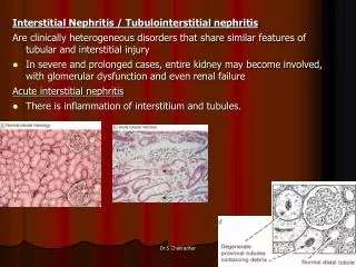
Tubulointristitial Nephritis A disorder of the interstitium and tubules
Acute Interstitial Nephritis 70% Drug hypersensitivity • 30% Antibiotics: PCNs (Methicillin), Cephalosporins, Cipro • Sulfa drugs • NSAIDs • Allopurinol... 15% Infection • Strep, Legionella, CMV, other bact/viruses 8% Idiopathic 6% Autoimmune Dz (Sarcoidosis, Tubulointerstitial nephritis/Uveitis)
AIN from Drugs Renal damage is NOT dose-dependent May take weeks after initial exposure to drug • More common is seen several months to a year after use But as early as 1 week after medication is begun • Fever (27%) • Serum Eosinophilia (23%) • Maculopapular rash (15%) • Bland sediment or WBCs, RBCs, non-nephrotic proteinuria • WBC Casts are pathognomonic! • Urine eosinophils on Wright’s or Hansel’s Stain • Also see urine eos in RPGN, renal atheroemboli • Leukocytoclastic vasculitis
Interstitial nephritis • Acute allergic IN- presents with fever, maculopapular rash, arthralgia, eosinophilia with use of certain drugs or systemic infection • UA_ microscopic hematuria, pyuria, non nephrotic proteinuria, eosinophiluria • Usually resolves after d/c of offending drug and steroids
Clinical Presentation Drug-Induced AIN AIN of any cause • Rash 15% • Fever 27% • Eosinophilia 23% • Triad 10% Nausea Vomiting Malaise
Laboratory Manifestions • Acute rise in plasma creatinine concentration • Eosinophilia and eosinophiluria • Urine sediment: wbcs, rbcs, white cell casts • Proteinuria (< 1 g/day) • Signs of tubulointerstitial damage
Acute bilateral pyelonephritis • Flank pain, toxic, febrile • U/A : pyoria, hematuria, proteinuria, bacteriuria • B/C & U/C help to diagnosis
Infiltrative/Autoimmune Causes of AIN Sarcoidosis Sjogren’s Syndrome TINU Leukemia Lymphoma Systemic lupus erythematosus
Renal biopsy Indications Uncertainty of diagnosis Advanced renal failure Lack of spontaneous recovery following removal of offending drug
Treatment Discontinuation of offending agent Corticosteroids • Prednisone 1 mg/kg to a max of 40-60 mg x 1-2 weeks • IV Methylprednisolone 0.5 – 1 g/day x 3 days
AIN Management • Remove offending agent • Most patients recover full kidney function in 1 year • Poor prognostic factors • ARF > 3 weeks • Advanced age at onset
Causes of chronic interstitial nephritis • Toxins( analgesic nephropathy, lead nephropathy) • Infection (chronic pyelonephritis) • Autoimmune( Sjogren syndrome, SLE, renal rejection) • Metabolic( hyperuricemia, hypercalcemia) • Radiation • Neoplastic infiltration( leukemia, lymphoma, multiple myeloma) • Hereditary renal diseases(ADPKD,MCD,MSK)
Analgesic abuse nephropathy • chronic interstitial nephritis • Result from excessive consumption (Phenacetin & Aspirin) • Dose dependent (at least 1 kg) • Being responsible for 1% to 3% of ESRD cases
The earliest lesion is thickening of GBM of vasa recta and capillary medulla then at later stage, focal area of necrosis in medulla and fibrosis and atrophy of tubules that leads to papillary necrosis and calcification. • Pathogenesis: phenacetin and its metabolites concentrate in the medulla where induce the generation of reactive metabolites • Aspirin potentiate the toxicity of phenacetin
Bacterial infection • bacterial infection of the renal parenchyma causes interstitial nephritis • infection without anatomical abnormality seldom produces permanent damage • obstruction (stones, prostate etc) in combination with infection can cause progressive disease • tuberculosis causes extensive destruction from granulomata, fibrosis and caseation
The upper and lower poles with compound papilla in ascending infection • At first, interstitial edema and PMN infiltration, then formation of irregular abscesses and eventually scars • Risk factors: diabetes mellitus obstruction delayed antimicrobial therapy severe infection with ATN reflux nephropathy papillary necrosis
Consequences of urinary tract obstruction • Reduced glomerular filtration rate • Reduced renal blood flow (after initial rise) • Impaired renal concentrating ability • Impaired distal tubular function • Nephrogenic diabetes insipidus • Renal salt wasting • Renal tubular acidosis • Impaired potassium concentration • Reduced RBF leads to renal ischemia tubular atrophy
baseline Hours Acute urinary tract obstruction Functional consequences
myeloma • Bence-Jones protein (light chains from malignant plasma cell clone) causes interstitial nephritis, tubular obstruction(cast nephropathy) and amyloid deposition • Myeloma kidney :the classic pathologic include THP+LC casts in dilated, atrophic distal tubuls with infiltration monocyte & macrophage & plasma cells that produce giant cells • LCs are nephrotoxic through direct injury of tubular epithelial cells & intrarenal obstruction from cast formation
Predisposing factors: LC concentration LC isoelectric point intraluminal PH tubular flow rate presence of Tamm-Horsfall Pr
Hypercalcemic Nephropathy • Chronic hypercalcemia is seen in Hyperparathyroidism Sarcoidosis Multiple myeloma Vitamin D toxicity Metastasis bone disease
Hypercalcemia decrease GFR through renal vasoconstriction • Calcium deposition in distal nephron and interstitial that leads to mononeuclear cell infiltration and tubular necrosis • Defective concentration ability, poliuria, nocturia • Nephrocalcinosis & nephrolitiasis
WBC Casts Cells in the cast have nuclei (unlike RBC casts) Pathognomonic for Acute Interstitial Nephritis
1. hyaline casts 2. muddy brown casts 3. RBCs 4. RBC casts 5. Oval fat bodies 6. eosinophils A. ATN B. prerenal azotemia C. glomerulonephritis D. nephrolithiasis E. interstitial disease F. nephrotic syndrome Match:
Question • 1. 65 year old with H/o HTN, ventricular arrythmias controlled on Amiodarone, OA on NSAIds presents with puffiness on face on waking up. Has bilateral pitting dema. UA 3+ prot, 3 RBC., 15-20 WBC • 24 h prot – 4 g/day • BUN/creat 80/5 • Serum albumin 2.8, TSH normal. The most likely diagnosis? • A) amiodarone induced hypothyroidism • B) RPGN • C) NSAIDs induced nephrotic sdr and interstitial nephritis