Download
1 / 23
230 likes | 354 Views
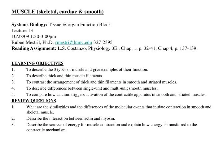
MUSCLE (skeletal, cardiac & smooth) Systems Biology: Tissue & organ Function Block Lecture 13 10/28/09 1:30-3:00pm Ruben Mestril, Ph.D: rmestri@lumc.edu 327-2395 Reading Assignment: L.S. Costanzo, Physiology 3E., Chap. 1, p. 32-41: Chap 4, p. 137-139. LEARNING OBJECTIVES

E N D
MUSCLE (skeletal, cardiac & smooth)Systems Biology: Tissue & organ Function BlockLecture 1310/28/09 1:30-3:00pmRuben Mestril, Ph.D: rmestri@lumc.edu 327-2395Reading Assignment:L.S. Costanzo, Physiology 3E., Chap. 1, p. 32-41: Chap 4, p. 137-139. • LEARNING OBJECTIVES • To describe the 3 types of muscle and give examples of their function. • To describe thick and thin muscle filaments. • To contrast the arrangement of thick and thin filaments in smooth and striated muscles. • To describe differences between single-unit and multi-unit smooth muscles. • To compare how calcium triggers activation of the contractile apparatus in smooth and striated muscles. • REVIEW QUESTIONS • What are the similarities and the differences of the molecular events that initiate contraction in smooth and skeletal muscle. • Describe the interaction between actin and myosin. • Describe the sources of energy for muscle contraction and explain how energy is transferred to the contractile mechanism.
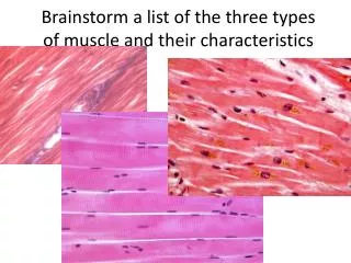
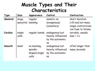
Three types of Muscle: Skeletal, Cardiac and Smooth muscle. • Function is to generate force or movement in response to a physiological stimulus. • Skeletal muscle: maintains contractile force for long periods. • Cardiac muscle: contracts for brief periods with each heartbeat, but for a lifetime. • Smooth muscle: must contract without fatigue for very long periods. Muscle Contraction: In all three muscle types contraction depends on rise in free Ca2+ concentration. Each muscle type has specialized plasma membrane, cytoskeleton, endoplasmic reticulum and metabolic pathways for energy generation and utilization.
MUSCLE FILAMENTS Thin filaments (actin, tropomyosin and troponin). Actin made up of filamentous or F-actin is closely associated to the actin binding proteins: tropomyosin and troponin. Thick filaments made up of multiple myosin-II molecules (hexamer: 2 heavy chains, 2 regulatory light chains and 2 alkali light chains). Heavy chains bind the actin of the thin filaments, alkali light chains stabilize myosin head regions and the regulatory light chain regulates the ATPase activity of myosin.
Figure 1-21 Structure of thick (A) and thin (B) filaments of skeletal muscle. Troponin is a complex of three proteins: I, troponin I; T, troponin T; and C, troponin C.
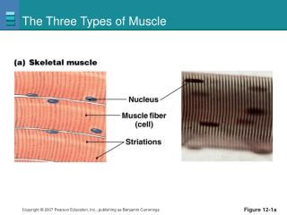
Muscle Contraction. Striated muscle cells are densely packed with myofibrils that contain ordered arrays of thick and thin filaments. Striated muscle (skeletal and cardiac muscle) where each myocyte or fiber contains myofibrils in the diameter of a Z-disk. Each myofibril is made up of sarcomeres composed of myofilaments, both thick filaments made up of myosin and thin filaments made up of actin. The A-bands mark the center of the sarcomere (containing the thick filaments) and are the area of cross-bridge formation between thin and thick filaments. The I-bands are outside of the A-band and contain the thin filaments.
Figure 1-22 Arrangement of thick and thin filaments of skeletal muscle in sarcomeres.
TRANSVERSE (T) TUBULES Transverse tubules are extensive network of muscle cell membrane or sarcolemmal membrane that are responsible for carrying depolarization from the action potentials at the muscle cell surface to the interior of the muscle fiber. The T tubules are in close contact with the sarcoplasmic reticulum that is the site of storage and release of Ca2+ during excitation-contraction coupling.
Figure 1-23 Transverse tubules and sarcoplasmic reticulum of skeletal muscle. The transverse tubules are continuous with the sarcolemmal membrane and invaginate deep into the muscle fiber, making contact with terminal cisternae of the sarcoplasmic reticulum.
Muscle cell excitation. Skeletal muscle contracts in response to neuromuscular synaptic transmission where one neuron innervates several skeletal muscle cells to form a “motor unit”. Transmission is through inotropic (nicotine) Ach receptors. Cardiac muscle contracts in response to the propagation of electrical signals from one cardiac cell to another across gap junctions. Electrical signals are generated in the pacemaker region of the heart (sinoatrial node), that generates action potentials transmitted from cell to cell through gap junctions. Chemical synapses only modulate but do not initiate cardiac contraction. In smooth muscle, neuromuscular transmission may initiate contraction or may just modulate contraction initiated by another mechanism.
Figure 1-24 Temporal sequence of events in excitation-contraction coupling in skeletal muscle. The muscle action potential precedes a rise in intracellular [Ca2+], which precedes contraction.
Excitation – Contraction Coupling • Invaginations of the sarcolemma facilitate communication between the surface of the cell and it’s interior. • In skeletal muscle, depolarization of the T-tubule membrane leads to Ca2+ release from the sarcoplasmic reticulum at the Triad. • In cardiac muscle, Ca2+ entry through L-type Ca2+ channels is amplified by Ca2+-induced Ca2+ release from the sarcoplasmic reticulum. • In smooth muscle, both extracellular and intracellular Ca2+ activate contraction. • Smooth muscle contraction may also occur independently of increases in [Ca2+]i. • Terminating Contraction • In skeletal, cardiac and smooth muscle, terminating contraction requires re-uptake of Ca2+ into the sarcoplasmic reticulum. • In smooth muscle, terminating contraction also requires dephosphorylation of the myosin light chain.
Figure 1-25 Cross-bridge cycle in skeletal muscle. Mechanism by which myosin "walks" toward the plus end of the actin filament. A-E, See the discussion in the text. ADP, Adenosine diphosphate; ATP, adenosine triphosphate; Pi, inorganic phosphate.
Regulating Muscle Contraction • Muscle contractions produce force and/or shortening and, in the extreme, can be studied under either isometric or isotonic conditions. • Muscle length influences tension development by determining the degree of overlap between actin and myosin filaments. • At high loads, the velocity of shortening is lower because more cross-bridges are simultaneously active. • In a single skeletal muscle fiber, the force developed may be increased by summing multiple twitches in time. • In a whole skeletal muscle, the force developed may be increased by summing the contraction of multiple fibers. • In cardiac muscle, increasing the entry of Ca2+ enhances the contractile force. • In smooth muscle, contractile force is enhanced by increasing the entry of Ca2+, as well as by increasing the Ca2+ sensitivity of the contractile apparatus. • Smooth muscle maintains high force at low energy consumption.
Figure 1-26 Length-tension relationship in skeletal muscle. Maximal active tension occurs at muscle lengths where there is maximal overlap of thick and thin filaments.
Figure 1-27 Initial velocity of shortening as a function of afterload in skeletal muscle.
EXCITATION-CONTRACTION COUPLING IN CARDIAC MUSCLE CELLS • Cardiac action potential is initiated in the myocardial cell membrane resulting in an inward Ca2+ current. • Entry of Ca2+ into the myocardial cell produces an increase in intracellular Ca2+ concentration that triggers release of more Ca2+ from stores in the SR (Ca2+ -induced Ca2+ release). • Ca2+ binds to troponin C so tropomyosin is moved out of the way and actin and myosin are able to interact. Actin and Moysin cross-bridges form and break permitting the thin and thick filaments move past each other producing tension. • The magnitude of the tension developed by myocardial cells is proportional to the intracellular Ca2+ concentration.
Figure 4-18 Excitation-contraction coupling in myocardial cells. See the text for an explanation of the circled numbers. SR, Sarcoplasmic reticulum.
In cardiac muscle, Ca2+ entry through L-type Ca2+ channels is amplified by Ca2+ -induced Ca2+ release from the sarcolasmic reticulum Donald Bers, Nature 415:198-205; 2002.
Smooth muscle may contract in response to either neuromuscular synaptic transmission or electrical coupling. • Smooth muscle may be formed into “multiunit smooth muscle” where smooth muscle cells are innervated by more than one neuron and there is little electrical coupling (gap junctions) between smooth muscle cells. Found mostly in the iris and ciliary body of the eye, the piloerector muscles of the skin and some blood vessels. • Other smooth muscles depend mainly on electrical coupling through gap junctions permitting coordinated contraction among cells to form the “unitary smooth muscle” found in the gastrointestinal tract, the uterus and many blood vessels (also called visceral smooth muscle).
Figure 1-28 Sequence of molecular events in contraction of smooth muscle. When myosin and actin bind, cross-bridges form and produce tension. When nonphosphorylated myosin and actin bind, latch-bridges form but do not cycle; a tonic level of tension then is produced. ADP, Adenosine diphosphate; ATP, adenosine triphosphate; Myosin-P, phosphorylated myosin; Pi, inorganic phosphate.
Action potentials of smooth muscle may be brief or prolonged. In unitary smooth muscle, action potentials maybe a simple spike, a spike followed by a plateau or a series of spikes on top of slow waves. Some of these smooth muscle cells can initiate spontaneous electrical activity. Some of this spontaneous electrical activity results in regular and repetitive oscillations referred as slow waves. In multiunit smooth muscle, action potentials usually do not occur.
Figure 1-29 Mechanisms for increasing intracellular [Ca2+] in smooth muscle. ATP, Adenosine triphosphate; G, GTP-binding protein (G protein); IP3, inositol 1,4,5-triphosphate; PIP2, phosphatidylinositol 4,5-diphosphate; PLC, phospholipase C; R, receptor for hormone or neurotransmitter.