Download
1 / 58
770 likes | 1.58k Views
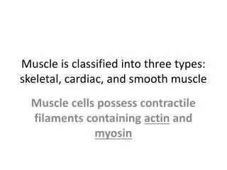
The Three Types of Muscle. Figure 12-1a. The Three Types of Muscle. Figure 12-1b. The Three Types of Muscle. Figure 12-1c. Skeletal Muscle. Usually attached to bones by tendons Contract only in response to signal from somatic motor neuron Flexor: brings bones together

E N D
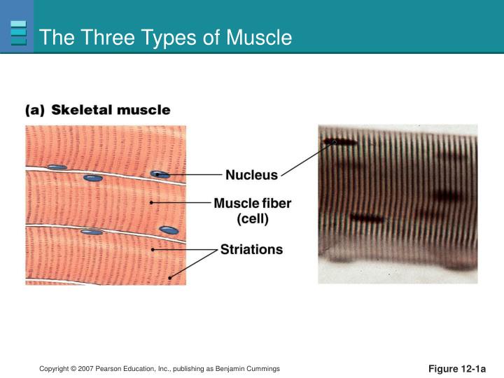
The Three Types of Muscle Figure 12-1a
The Three Types of Muscle Figure 12-1b
The Three Types of Muscle Figure 12-1c
Skeletal Muscle • Usually attached to bones by tendons • Contract only in response to signal from somatic motor neuron • Flexor: brings bones together • Extensor: bones move away • Antagonistic muscle groups: flexor-extensor pairs
Antagonistic Muscle Groups Figure 12-2a
Antagonistic Muscle Groups Figure 12-2b
Anatomy Summary: Skeletal Muscle Figure 12-3a (1 of 2)
Anatomy Summary: Skeletal Muscle Figure 12-3a (2 of 2)
T-tubules and the Sarcoplasmic Reticulum Figure 12-4
Ultrastructure of Muscle Figure 12-3b
Ultrastructure of Muscle Myosin are motor proteins. 250 myosins join to form the thick filaments Figure 12-3e
Thin Filaments • Thin filaments are actin chains and regulatory proteins
Ultrastructure of Muscle Actin and myosin form crossbridges Under light microscope one sees repeating light & dark bands One repeat pattern is a “sarcomere” Z disks: attachment site for thin filaments I band: lightest band, region only occupied by thin filaments A band: darkest, entire length of thick filaments, overlap with thin filaments H zone: Thick filaments only (center of A band) M line: attachment site for thick filaments Figure 12-3d
Ultrastructure of Muscle Sarcomere A band (c) Z disk Z disk Myofibril M line I band H zone Figure 12-3c
Ultrastructure of Muscle Sarcomere A band (c) Z disk Z disk Myofibril M line I band H zone (d) Titin Z disk Z disk M line Figure 12-3c–d (1 of 2)
Ultrastructure of Muscle Sarcomere A band (c) Z disk Z disk Myofibril M line I band H zone (d) Titin Z disk Z disk M line M line Thick filaments Figure 12-3c–d (2 of 2)
Ultrastructure of Muscle Sarcomere A band (c) Z disk Z disk Myofibril M line I band H zone (d) Titin Z disk Z disk M line M line Thick filaments (e) Myosin heads Hinge region Myosin tail Myosin molecule Figure 12-3c–e (1 of 2)
Ultrastructure of Muscle Sarcomere A band (c) Z disk Z disk Myofibril M line I band H zone (d) Titin Z disk Z disk M line M line Thin filaments Thick filaments Titin (e) Myosin heads Hinge region Myosin tail Myosin molecule Figure 12-3c–e (2 of 2)
Ultrastructure of Muscle Sarcomere A band (c) Z disk Z disk Myofibril M line I band H zone (d) Titin Z disk Z disk M line M line Thin filaments Thick filaments Titin (f) Troponin Nebulin (e) Myosin heads Hinge region Myosin tail Tropomyosin G-actin molecule Actin chain Myosin molecule Figure 12-3c–f
Titin and Nebulin Figure 12-6
Muscle Contraction • Muscle tension: force created by muscle • Load: weight that opposes contraction • Contraction: creation of tension in muscle • Relaxation: release of tension
Summary of Muscle Contraction Figure 12-7
Changes in Sarcomere Length during Contraction Figure 12-8
The Molecular Basis of Contraction Myosin filament Tight binding in the rigor state. The crossbridge is at a 45° angle relative to the filaments. ATP binds to its binding site on the myosin. Myosin then dissociates from actin. 45° Myosin binding sites 1 2 ATP binding site 3 2 4 1 G-actin molecule ADP ATP 3 2 4 1 3 2 1 4 5 At the end of the power stroke, the myosin head releases ADP and resumes the tightly bound rigor state. 6 The ATPase activity of myosin hydrolyzes the ATP. ADP and Pi remain bound to myosin. 3 ADP Contraction- relaxation Pi Pi Sliding filament 3 2 4 1 3 2 4 1 5 Actin filament moves toward M line. 90° Pi 3 2 4 1 Release of Pi initiates the power stroke. The myosin head rotates on its hinge, pushing the actin filament past it. 5 The myosin head swings over and binds weakly to a new actin molecule. The crossbridge is now at 90º relative to the filaments. 4 Figure 12-9
The Molecular Basis of Contraction Tight binding in the rigor state. The crossbridge is at a 45° angle relative to the filaments. 1 Myosin filament 45 ° ATP binding site Myosin binding sites 3 2 4 1 G-actin molecule Figure 12-9, step 1
The Molecular Basis of Contraction Tight binding in the rigor state. The crossbridge is at a 45° angle relative to the filaments. ATP binds to its binding site on the myosin. Myosin then dissociates from actin. 1 2 Myosin filament 45 ° ATP binding site Myosin binding sites ATP 3 2 4 3 2 1 4 1 G-actin molecule Figure 12-9, steps 1–2
The Molecular Basis of Contraction The ATPase activity of myosin hydrolyzes the ATP. ADP and Pi remain bound to myosin. 3 ADP Pi 3 2 4 1 Figure 12-9, step 3
The Molecular Basis of Contraction The ATPase activity of myosin hydrolyzes the ATP. ADP and Pi remain bound to myosin. The myosin head swings over and binds weakly to a new actin molecule. The cross- bridge is now at 90º relative to the filaments. 3 4 ADP 90° Pi Pi 3 2 4 1 3 2 4 1 Figure 12-9, steps 3–4
The Molecular Basis of Contraction Release of Pi initiates the power stroke. The myosin head rotates on its hinge, pushing the actin filament past it. 5 Pi 3 2 4 1 5 Actin filament moves toward M line. Figure 12-9, step 5
The Molecular Basis of Contraction Release of Pi initiates the power stroke. The myosin head rotates on its hinge, pushing the actin filament past it. At the end of the power stroke, the myosin head releases ADP and resumes the tightly bound rigor state. 5 6 ADP Pi 3 2 3 2 4 1 4 1 5 5 Actin filament moves toward M line. Figure 12-9, steps 5–6
The Molecular Basis of Contraction Myosin filament Tight binding in the rigor state. The crossbridge is at a 45° angle relative to the filaments. ATP binds to its binding site on the myosin. Myosin then dissociates from actin. 45° 1 2 ATP binding site 3 2 4 1 G-actin molecule ADP ATP 3 2 4 1 3 2 1 4 5 At the end of the power stroke, the myosin head releases ADP and resumes the tightly bound rigor state. 6 The ATPase activity of myosin hydrolyzes the ATP. ADP and Pi remain bound to myosin. 3 ADP Contraction- relaxation Pi Pi Sliding filament 3 2 4 1 3 2 4 1 5 Actin filament moves toward M line. 90° Pi 3 2 4 1 Release of Pi initiates the power stroke. The myosin head rotates on its hinge, pushing the actin filament past it. 5 The myosin head swings over and binds weakly to a new actin molecule. The crossbridge is now at 90º relative to the filaments. 4 Myosin binding sites Figure 12-9
In vitro assay of myosin-based motility Gliding assay Fluorescent actin filaments Coverslip coated with myosin Add ATP
Optical tweezers (laser optical trap) • Can measure single steps of molecular motors 3 pN force generated Measure step size in nm
X-ray crystal structures In the beginning of the movie, the myosin heads are in the prestroke ADP-Pi state (yellow) and the catalytic cores bind weakly to actin. Once a head docks properly onto an actin subunit (green), phosphate (Pi) is released from the active site. Phosphate release increases the affinity of the myosin head for actin and swings the converter/lever arm to the poststroke, ADP state (transition from yellow to red). The swing of the lever arm moves the actin filament by ~100 Å; the exact distance may vary from cycle to cycle depending upon the initial prestroke binding configuration of the myosin on actin. After completing the stroke, ADP dissociates and ATP binds to the empty active site, which causes the catalytic core to detach from actin. The lever arm then recocks back to its prestroke state (transition from red to yellow).
Regulatory Role of Tropomyosin and Troponin Figure 12-10a
Regulatory Role of Tropomyosin and Troponin (b) Initiation of contraction Ca2+ levels increase in cytosol. 1 ADP 1 Cytosolic Ca2+ Figure 12-10b, step 1
Regulatory Role of Tropomyosin and Troponin (b) Initiation of contraction Ca2+ levels increase in cytosol. 1 Ca2+ binds to troponin. 2 ADP TN 2 1 Cytosolic Ca2+ Figure 12-10b, steps 1–2
Regulatory Role of Tropomyosin and Troponin (b) Initiation of contraction Ca2+ levels increase in cytosol. 1 Ca2+ binds to troponin. 2 3 Tropomyosin shifts, exposing binding site on G-actin ADP Troponin-Ca2+ complex pulls tropomyosin away from G-actin binding site. 3 TN 2 1 Cytosolic Ca2+ Figure 12-10b, steps 1–3
Regulatory Role of Tropomyosin and Troponin (b) Initiation of contraction Ca2+ levels increase in cytosol. 1 4 Power stroke Ca2+ binds to troponin. 2 3 Tropomyosin shifts, exposing binding site on G-actin ADP Troponin-Ca2+ complex pulls tropomyosin away from G-actin binding site. 3 TN Myosin binds to actin and completes power stroke. 4 2 1 Cytosolic Ca2+ Figure 12-10b. Steps 1–4
Regulatory Role of Tropomyosin and Troponin (b) Initiation of contraction Ca2+ levels increase in cytosol. 1 4 Power stroke Ca2+ binds to troponin. 2 3 Tropomyosin shifts, exposing binding site on G-actin Pi ADP Troponin-Ca2+ complex pulls tropomyosin away from G-actin binding site. 3 TN Myosin binds to actin and completes power stroke. 4 5 2 G-actin moves Actin filament moves. 5 1 Cytosolic Ca2+ Figure 12-10b, steps 1–5
Excitation-Contraction Coupling Somatic motor neuron releases ACh at neuro- muscular junction. 1 (a) 1 Axon terminal of somatic motor neuron ACh Muscle fiber Motor end plate Sarcoplasmic reticulum T-tubule Ca2+ DHP receptor Tropomyosin Z disk Troponin Actin M line Myosin head Myosin thick filament Figure 12-11a, step 1
Excitation-Contraction Coupling Somatic motor neuron releases ACh at neuro- muscular junction. Net entry of Na+ through ACh receptor-channel initiates a muscle action potential. 1 2 (a) 1 Axon terminal of somatic motor neuron ACh Muscle fiber potential Action K+ Action potential 2 Na+ Motor end plate Sarcoplasmic reticulum T-tubule Ca2+ DHP receptor Tropomyosin Z disk Troponin Actin M line Myosin head Myosin thick filament Figure 12-11a, steps 1–2
Excitation-Contraction Coupling Action potential in t-tubule alters conformation of DHP receptor. 3 (b) 3 Ca2+ Figure 12-11b, step 3
Excitation-Contraction Coupling Action potential in t-tubule alters conformation of DHP receptor. DHP receptor opens Ca2+ release channels in sarcoplasmic reticulum and Ca2+ enters cytoplasm. 3 4 (b) 4 3 Ca2+ Ca2+ released Figure 12-11b, steps 3–4
Excitation-Contraction Coupling Action potential in t-tubule alters conformation of DHP receptor. DHP receptor opens Ca2+ release channels in sarcoplasmic reticulum and Ca2+ enters cytoplasm. 3 4 5 Ca2+ binds to troponin, allowing strong actin- myosin binding. (b) 4 3 Ca2+ Ca2+ released 5 Figure 12-11b, steps 3–5
Excitation-Contraction Coupling Action potential in t-tubule alters conformation of DHP receptor. DHP receptor opens Ca2+ release channels in sarcoplasmic reticulum and Ca2+ enters cytoplasm. 3 4 5 Ca2+ binds to troponin, allowing strong actin- myosin binding. (b) 4 3 Ca2+ Ca2+ released 5 6 Myosin thick filament M line Myosin heads execute power stroke. 6 Figure 12-11b, steps 3–6
Excitation-Contraction Coupling Action potential in t-tubule alters conformation of DHP receptor. DHP receptor opens Ca2+ release channels in sarcoplasmic reticulum and Ca2+ enters cytoplasm. 3 4 5 Ca2+ binds to troponin, allowing strong actin- myosin binding. (b) 4 3 Ca2+ Ca2+ released 5 7 6 Myosin thick filament M line Distance actin moves Actin filament slides toward center of sarcomere. Myosin heads execute power stroke. 6 7 Figure 12-11b, steps 3–7
Electrical and Mechanical Events in Muscle Contraction A twitch is a single contraction-relaxation cycle Figure 12-12
Phosphocreatine Provides ATP at beginning of exercise needed for contraction Figure 12-13
Muscle Fatigue Locations and possible causes of muscle fatigue Figure 12-14