Download
1 / 34
340 likes | 345 Views
NERVOUS SYSTEM. It is the master controlling and communicating system of the body. Structurally , it has two subdivisions : (1) Central nervous system. (2) Peripheral nervous system. NERVOUS SYSTEM. Functionally , it is divided into : Somatic N.S . : It controls voluntary activities.

E N D
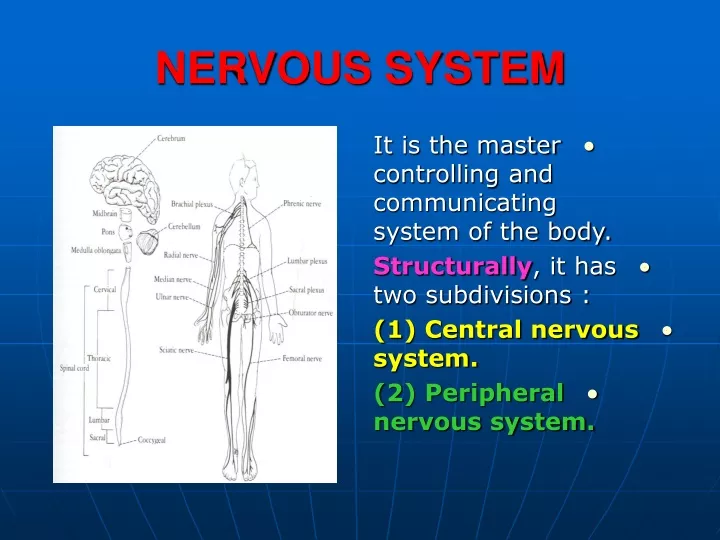
NERVOUS SYSTEM • It is the master controlling and communicating system of the body. • Structurally, it has two subdivisions : • (1) Central nervous system. • (2) Peripheral nervous system.
NERVOUS SYSTEM • Functionally, it is divided into : • Somatic N.S. : It controls voluntary activities. • Autonomic N.S. :It controls involuntary activities.
CENTRAL NERVOUS SYSTEM • It is composed of the brain and spinal cord. • They occupy the dorsal body cavity. • They act as the integrating and command centers of the nervous system.
CENTRAL NERVOUS SYSTEM • Its interior is organized into Gray and White matter.
CENTRAL NERVOUS SYSTEM • Gray matter: • Consists of nerve cells (neurons) embedded in neuroglia. • Each neuron has two types of nerve processes: • Dendrites : short processes of the cell body. • Axon : longest process of the cell body.
CENTRAL NERVOUS SYSTEM • White matter: • Consists of nervefibers embedded in neuroglia.
PERIPHERAL NERVOUS SYSTEM • It is the part of the nervous system outside the CNS. • It consists of (12) paircranial and (31) pair ofspinal nerves. • These nerves act as communication lines between all parts of the body and the CNS.
PERIPHERAL NERVOUS SYSTEM • The nerves are formed of grayish white cords. • Each cord is formed of nerve fibers (axons) supported by delicate areolar tissue.
CRANIAL NERVES • The twelve pair of nerves supply structures in the head and neck. • The 10th(vagus) supplies structures in the thorax and abdomen.
SPINAL NERVES • They are named according to the region of the spinal cord to which they are associated. • They are: 8)cervical.12)thoracic.5)lumbar. • 5)sacraland • (1)coccygeal.
LENGTH OF SPINAL NERVES • In the upper cervical region, they are short and horizontal. • The lumbar and sacral nerves below the level of termination, form a vertical bundle (Cauda Equina).
LEVEL OF TERMINATION OF S.C. • Adults : • It terminates inferiorly at the lower border of the 1st lumbar vertebra. • Children : • It is relatively longer and ends at the upper border of the third lumbar vertebra.
SPINAL NERVES • Each spinal nerve is attached to the spinal cord by two roots : Anterior and Posterior.
ANTERIOR ROOT • It carries nerve impulses away from the central nervous system (Efferent) fibers. • The efferent fibers to the skeletal muscles are called (Motor) fibers. • They arise from the anterior gray horn cells of the spinal cord
POSTERIOR ROOT • It carries impulses to the central nervous system (Afferent) fibers. • It conveys sensations of pain, touch and temperature (Sensory) fibers. • Their cell bodies are located in the posterior root ganglion.
SPINAL NERVE • At the intervertebral foramen the anterior and posterior roots unite to form a spinal nerve. • Each spinal nerve is a mixture of motor and sensory fibers.
FORMATION OF THE RAMI • On emerging from the foramen, each spinal nerve divides into a large ventral (anterior) ramus and a smaller dorsal (posterior) ramus.
DISTRIBUTION OF THE RAMI • The posterior rami pass posteriorly to supply the skin and muscles of the back. • The anterior rami supply the skin and muscles of the limbs and the anterolateral wall of the body.
PLEXUSES • At the root of the upper limb, the anterior rami unite to form the Cervical and Brachial plexuses. • At the root of the lower limb, they form the Lumbar and Sacral plexuses.
AUTONOMIC NERVOUS SYSTEM • It is concerned with the innervation of the involuntary structures (heart, smooth muscles ,glands). • It is divided into :
AUTONOMIC NERVOUS SYSTEM • 1. Sympatheticsystem: it prepares the body for an emergency. • 2. Parasympathetic system : • It conserves and restores energy. • Each system has : • Efferent, Afferent fibers and ganglia to relay.
SYMPATHETIC SYSTEM • Efferent fibers : • Their cell bodies are located in the lateral column of the spinal cord from the level of (T1- L2). • They are divided into :
SYMPATHETIC SYSTEM • A. preganglionic fibers • They leave the lateral column of the spinal cord in the anterior nerve roots. • They pass in the White ramicommunicantes to the paravertebral ganglia of the sympathetic trunk.
SYMPATHETIC TRUNKS • They are two ganglionated trunks that extend close to the vertebral column. • Each trunk is formed of (3) ganglia in the neck, (12) in the thorax, (5) in the lumbar and (5) in the sacral. • The two trunks end inferiorly by forming one ganglion. • Ganglion : collection of nerve cells (neurons) outside the central nervous system.
PREGANGLIONIC FIBERS • 1. They relay at the corresponding ganglia by synapsing with an excitor cell of the ganglion. • 2. Fibers in the upperthorax may travel up in the sympathetic trunk to relay in the cervical ganglia.
PREGANGLIONIC FIBERS • 3. Splanchnic nerves • They pass through the thoracic sympathetic ganglia withoutrelay. • They pass through the diaphragm. • They are : • a. Greater Splanchnic nerve (5th -9th ) thoracic ganglia. • It relays in the celiac ganglia.
PREGANGLIONIC FIBERS • b. Lesser splanchnic nerve (10th & 11th ) ganglia :it synapses with ganglia in the lower part of the celiac trunk. • C. Least splanchnic nerve (12th) ganglion : • It relays in ganglia of the renal plexus.
POSTGANGLIONIC FIBERS • They pass through the Gray rami communicantes to join the spinal nerves to supply the smooth muscles in the blood vessels, sweat glands and erector pili muscles of the skin.
POSTGANGLIONIC FIBERS • The postganglionic fibers of the splanchnic nerves supply the smooth muscle and glands of the viscera.
AFFERENT FIBERS • They arise from the wall of the viscera. • They pass through the sympathetic ganglia without relay. • They enter the spinal nerve through the white rami. • They relay in the posterior root ganglion of the corresponding spinal nerve. • Their central axons pass to the spinal cord.
PARASYMPATHETIC SYSTEM • Efferent fibers(Preganglionic) : • (1) Cranial part : • Their cells are located in the brain. • They form part of the cranial nerves (111, V11,1X &X).
PARASYMPATHETIC GANGLIA • They relay into ganglia situated close to the tructures they supply. • In the head & neck, they are the (ciliary, otic, pterygopalatine and submandibular) ganglia.
PARASYMPATHETIC SYSTEM • (2) Sacral part : • The fibers arise from the gray matter of the 2nd,3rd and 4thsacral spinal segments. • They form the Pelvic splanchnic nerves. • They relay in ganglia in the walls of the viscera or in the hypogastric plexuses.
PARASYMPATHETIC SYSTEM • Afferent fibers : • They pass from the viscera to the sensory ganglia of the cranial nerves or the posterior root ganglia of the sacrospinal nerves. • The central axons pass to the central nervous system.