Download
1 / 25
250 likes | 588 Views
Why I chose this paper?. Clinical/ radiological uncertainty of optimal diagnostic work-up during DGH attachmentImportant clinical/radiological problemVTE leading cause of maternal mortality and clinical diagnosis unreliableFinite risk of both radiological investigation and anticoagulant treatment

E N D
1. Diagnosis of suspected venous thromboembolic disease in pregnancy. AF Scarsbrook, AL Evans, AR Owen, FV Gleeson. Clinical Radiology (2006) 61, 1-12
Martin Hawkings
14th Feb 2006
2. Why I chose this paper? Clinical/ radiological uncertainty of optimal diagnostic work-up during DGH attachment
Important clinical/radiological problem
VTE leading cause of maternal mortality and clinical diagnosis unreliable
Finite risk of both radiological investigation and anticoagulant treatment
Medico-legal and no published guidelines
3. Approach Medline/Pubmed search limited to overview and/or systematic reviews
Discussion with consultant colleagues and evidence base required to inform local protocol development/ decision-making
4. Did the review address an important clinical question? �review the spectrum of diagnostic tests used in the investigation of suspected venous thromboembolic disease (VTE) in pregnancy; highlight the potential risks and benefits to mother and fetus of each of these tests; discuss the �albeit limited� guidelines for diagnosis of VTE in pregnancy; and suggest an appropriate imaging algorithm based on the available evidence.�
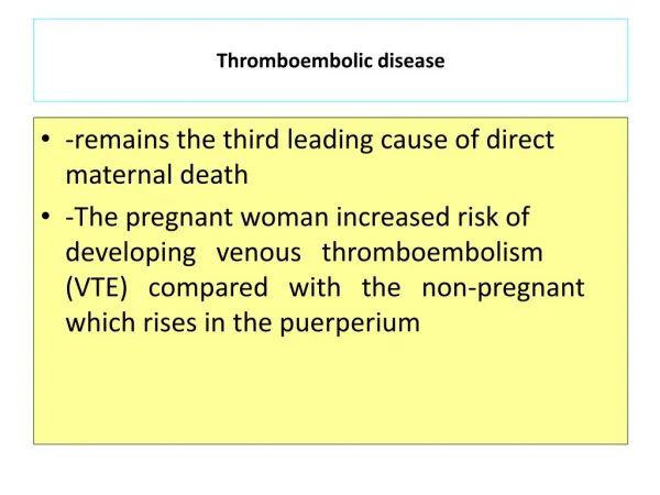
5. Background 2-4 fold increased risk VTE in pregnancy
Leading cause of maternal mortality
33% of maternal deaths; 50% first trimester
Incidence 1 in 2000 pregnancies
BTS guidelines for suspected acute PE advocates CTPA but does not include pregnant patients (Thorax 2003)
Older published guidelines addressing VTE in pregnancy do not include CTPA
6. Pathophysiology Pregnancy associated hypercoagulable state:
Progesterone induced ? venous compliance
Venous dilatation and ? flow
? procoagulants, ? natural anticoagulants
Associated risk factors in 2/3 of deaths
e.g. maternal age, weight and smoking, multi-parity, gestational diabetes, previous VTE and prolonged bed-rest
7. Radiological tests reviewed Chest X-ray
Radionuclide scintigraphy
CT pulmonary angiography (CPA)
Venography lower limb veins
Ultrasonography lower limb veins
Pulmonary angiography
Others
8. Was a thorough search done of appropriate databases/ sources? No statement of search methods, database used or inclusion/ exclusion criteria
Authors state that: �no large scale trials have been performed of VTE in pregnancy and recommendations empirical based on extrapolation from non-pregnant, small observational studies & personal experience
9. CXR Normal in 50% pregnant patients with PE
Exclude other causes pulmonary symptoms:
pneumonia; pneumothorax; lobar collapse
Abnormal findings due to PE:
focal opacities � atelectasis
effusion � regional oligaemia
pulmonary oedema (rare)
10. Venous ultrasonography Duplex scanning - primary diagnostic test DVT
symptomatic proximal sensitivity 97%; specificity 94%
distal sensitivity 11-100%; specificity 90-100%
NPV of serial US & cost-effectiveness of single negative demonstrated in non-pregnant patients
Prevalence lower limb DVT variable (13-93%) in proven PE - not validated as single diagnostic test
Concern of more frequent propagation/ risk of isolated pelvic thrombus and PE in pregnancy
11. Radionuclide diagnosis of VTE Well established non-invasive test in 2 large multi-centre prospective trials (PIOPED/ PISA-PED) but excluded pregnant patients
For �clinically significant PE�
High exclusion value of normal
High PPV of high probability exam
PE present in 30% non-diagnostic scintograms
Others validated negative serial ultrasound in low/ intermediate in low pre-test probability non-pregnant patients
12. Radionuclide in Pregnancy Recent small studies in pregnancy show different results compared to PIOPED:
normal 70% (v 10-30%)
low/ intermediate probability 25% (v 50-70%)
high probability 2% (v 10-15%)
Threshold effect of referral
Small studies questionable validity of NPV in normal/ intermediate in pregnancy
13. CTPA -new gold standard? Single slice CTPA comparable to PA for main, lobar and segmental thrombi:
sensitivity 90% � specificity 90%
Sub-segmental identification with multislice CTPA is being addressed in PIOPED II
Non-pregnant 3/12 test -ve recurrence
CTPA 1.1%; PA 0.9%; VQ 0.5%
In pregnancy dose reduction methods not standardised & may reduce accuracy
14. Pulmonary angiography Conventional gold standard invasive
Procedural mortality 0.5% from PIOPED
Radiation dose significantly higher CTPA
No more accurate than modern CTPA and not indicated in pregnancy
15. Fetal risk Fetal risk from diagnostic radiology low:
Threshold for deterministic effects - 50mGy
Excess malignancy up to 15 yrs of age after in-utero exposure - 1 in 16,000 per mSv
Many studies report doses � NRPB
Combined CXR, VQ, CTPA & PA fetal dose less than gestational background dose
Low risk from contrast (maternal &fetal)
16. Maternal risk Radiation dose in reducing dose order
PA ? msCTPA ? ssCTPA ? VQ
1st line CTPA may reduce sequential tests & overall dose but low NPPV in pregnancy
Organ-specific dose rate to breast in CTPA
7.4 mSv (5.5 - 13.1)
10 mGy increases breast cancer risk 10%
Low risk from iodinated contrast
17. Radiation and contrast risk to fetus and mother (NRPB)
18. Other tests CT venography - with CTPA increases sensitivity but contra-indicated in pregnancy
MRI/MRA potential for the future
Fast Imaging Steady State Precession (FISP)
MR direct thrombus imaging (MR-DTI)
safety/ efficacy not established in pregnancy and not widely available
19. DVT in Pregnancy Algorithm