Download
1 / 30
320 likes | 885 Views
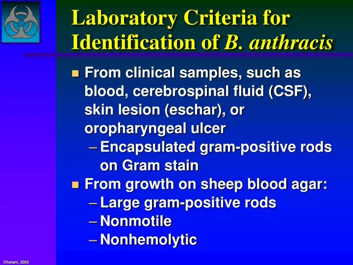
Laboratory Criteria for Identification of B. anthracis. From clinical samples, such as blood, cerebrospinal fluid (CSF), skin lesion (eschar), or oropharyngeal ulcer Encapsulated gram-positive rods on Gram stain From growth on sheep blood agar: Large gram-positive rods Nonmotile

E N D
Laboratory Criteria for Identification of B. anthracis • From clinical samples, such as blood, cerebrospinal fluid (CSF), skin lesion (eschar), or oropharyngeal ulcer • Encapsulated gram-positive rods on Gram stain • From growth on sheep blood agar: • Large gram-positive rods • Nonmotile • Nonhemolytic
Laboratory Criteria for Identification of B. anthracis • Rapid screening assay (PCR- and antigen-detection based) for use on cultures and directly on clinical specimens • Confirmatory criteria for identification of B. anthracis • Capsule production • Lysis by gamma-phage • Direct fluorescent antibody assay (DFA)
Gram Stain Morphologyof B. anthracis • Broad, gram-positive rod: 1–1.5 x 3–5 μ • Oval, central to subterminal spores: 1 x 1.5 μ with no significant swelling of cell • Spores usually NOT present in clinical specimens unless exposed to atmospheric O2
Gram-positive, spore-forming, non-motile bacillus B. anthracis
Gram Stain of Blood in Culture MediaGram-positive bacilli in long chains (original magnification 20). Enlargement shows typical "jointed bamboo-rod" appearance of Bacillus anthracis (original magnification 100). Reprinted from Borio et al.36
Gram stain Capsule production B. anthracis: Presumptive Identification Clinicalspecimen (blood, CSF, etc.) Isolate on SBA Colony morphology Hemolysis Motility Spores Gram stain Malachite green
Phage lysis Capsule DFA Capsule antigen Cell wall Horse blood Bicarbonate media (M’Fadyean stain India ink stain) (M’Fadyean Stain) B. anthracis: Confirmatory Identification Isolate
Immune Protection Against Anthrax • Live cellular vaccines • "Sterne" type live spore (toxigenic, non-capsulating) • Former USSR STI live spore (toxigenic, non-capsulating) • "Pasteur" type (mixed culture, reduced virulence) • Sterile, acellular vaccines • US "anthrax vaccine adsorbed" (AVA)—not licensed for use in civilian populations • UK "anthrax vaccine precipitated" (AVP) • Recombinant PA research vaccines • AI3+; Freund’s; Saponin, Monophosphoryl lipid A; Ribi
Anthrax in the US - 2001 • In October 2001, the first inhalational anthrax case in the United States since 1976 was identified in a media company worker in Florida. • A national investigation was initiated to identify additional cases and determine possible exposures to Bacillus anthracis. • Surveillance was enhanced through health-care facilities, laboratories, and other means to identify cases, which were defined as clinically compatible illness with laboratory-confirmed B. anthracis infection. • From October 4 to November 20, 2001, 22 cases of anthrax (11 inhalational, 11 cutaneous) were identified; 5 of the inhalational cases were fatal.
Epidemic curve for 22 cases of bioterrorism-related anthrax, United States, 2001.
Anthrax Meningitis • Hemorrhagic meningitis is a potential complication of anthrax, and may accompany cutaneous, gastrointestinal, or inhalational anthrax (seen in 50% of inhalational cases) • Death almost universal within 1-6 days after onset of illness • Rare survivors have been treated using appropriate antibiotics in combination with antitoxin, prednisone or both
Recommended Postexposure Prophylaxis to Prevent Inhalational Anthrax Initial Therapy Duration Adults Ciprofloxacin 60 days (including pregnant 500 mg PO BID women and OR immunocompromised) Doxycycline 100 mg PO BID Children Ciprofloxacin* 60 days 10–15 mg/kg PO Q 12 hrs Change to OR amoxicillin Doxycycline: if susceptible >8 yrs and >45 kg: 100 mg PO BID >8 yrs and <45 kg: 2.2 mg/kg PO BID <8 yrs: 2.2 mg/kg PO BID
Cutaneous Anthrax Treatment Protocol* for Cases Associated with Bioterrorist Events Category Initial Therapy (Oral) Duration Adults Ciprofloxacin 60 daysw (Including pregnant women 500 mg BID and immunocompromised) OR Doxycycline 100 mg BID Children Ciprofloxacin** 60 daysw (including immuno- 10–15 mg/kg Q 12 hrs compromised) OR Doxycycline: >8 yrs and >45 kg: 100 mg BID >8 yrs and <45 kg: 2.2 mg/kg BID <8 yrs: 2.2 mg/kg BID
Inhalational Anthrax Treatment Protocol* for Cases Associated with Bioterrorist Events Category Initial therapy (intravenous) Duration Adults Ciprofloxacin Switch to oral (Including pregnant 400 mg Q 12 hrs therapy when women** and OR clinically immunocompromised) Doxycycline appropriate: 100 mg Q 12 hrs Ciprofloxacin 500 mg BID ANDOR One or two additional Doxycycline 100 mg BID antimicrobials Continue for 60 days (IV and PO combined)
Smallpox • Worst-case scenario biological agent • Highly contagious once rash present (not before) • World’s population is largely susceptible • Up to 30% case fatality rate in non-immune • Secondary attack rate of 25-40% (10-20 secondary cases can be expected per index case) • No specific treatment available • Globally very few physicians currently practicing have seen actual cases • Virus has been weaponized by Soviets, uncertain who exactly owns viable stocks
Smallpox • Caused by Variola virus (Orthopox virus) • Immunization of U.S. civilian population suspended in 1980, U.S. military recruits in 1989 • Virus is stable in environment • Spread primarily by respiratory droplets, also by contact, fomites • Two distinct types of smallpox: • Variola Minor (Alastrim): diminutive lesions and mild systemic toxicity • Variola Major: Ordinary (subtypes discrete, semi-confluent, confluent), Modified, Flat, Hemorrhagic
Smallpox Clinical Presentation • 7-17 day incubation period • Prodromal phase: 2-4 days of malaise, fever, rigors, headache, backache, delirium • Rash then develops on face, hands, forearms and legs, including palms and soles (centrifugal distribution is important distinguishing feature). • Initial rash is maculopapular. In 1-2 days, lesions become vesicular, then evolve into round, tense pustules deeply imbedded in the dermis. Crusts form on 8th to 9th day of rash • Crusts separate to form depressed, hypopigmented scars
Smallpox Outbreak, Meschede Hospital, Germany, 1970 Wehrle PF, Bull WHO 1970;43:669
Diagnosis of Smallpox • Recognition of clinical features in early index cases is key • Identification of a single case of smallpox constitutes an international medical emergency, and should be considered evidence of a bioterrorist attack • Confirmation of diagnosis is made by demonstration of characteristic virions on electron microscopy of vesicular scrapings
Management of Smallpox • Immediate quarantine of affected and exposed individuals for 17 days • Only supportive care is available. Cidofovir has demonstrated in vitro activity • Immediate vaccination of all exposed persons with Vaccinia virus vaccine by inoculation with a bifurcated needle (scarification) • Administration of Vaccinia Immune Globulin (VIG), 0.6 ml/kg intramuscularly, concomitant with vaccination • Active and passive immunization is effective at preventing disease and death if given within 7 days of exposure
For the past half-century the main concern was a nuclear Armageddon caused by atomic weapons. However, with the discovery of various offensive biological warfare programs around the globe, the concern regarding intentional use of viruses and microorganisms as weapons of mass destruction increased during the past decade. The threat of biological warfare is real. It is now widely acknowledged that the biological weapons, in terms of destructiveness and in the generation of panic and civil disorder could produce an effect that is equivalent to that of a nuclear weapon. Chotani, 2003
Medical professionals globally should make sure that the world does not suffer a catastrophe as a result of the use of biowarfare pathogens Chotani, 2003
War on Diseases • The recent emphasis on bioterrorism: important truth about infectious diseases • Even without the element of intentional terror, diseases are a huge source of human suffering—and a tremendously destabilizing force • Nearly half of the world’s premature deaths (deaths under the age of 45) are caused by infectious diseases • Some 30 million infants in developing countries remain unprotected by the lifesaving childhood vaccines that in the rest of the world are administered routinely; a million die each year from measles alone
Pathogens of BT do threaten humanity but let us NOT loose sight of the naturally occurring microbes that threaten billions of people in a world weakened by poverty, war, lack of clean water, and inattention specially during wars & complex humanitarian emergencies Chotani, 2003