Download
1 / 39
400 likes | 606 Views
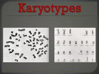
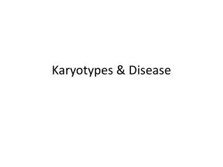
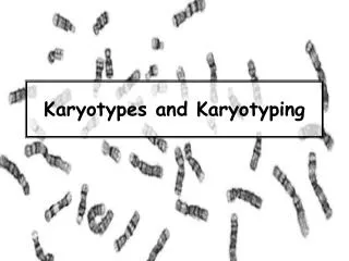
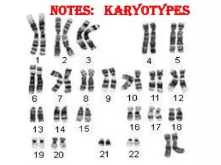
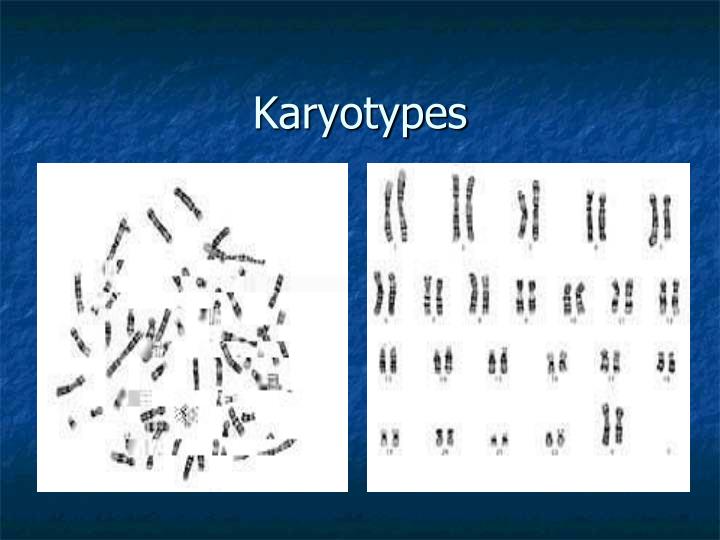
Karyotypes. Karyotype. A picture of the chromosomes from a human cell arranged in pairs by size First 22 pairs are called autosomes Last pair are the sex chromosomes XX female or XY male. Karyotype Procedure. 5 ml of blood is removed from the patient.

E N D
Karyotype • A picture of the chromosomes from a human cell arranged in pairs by size • First 22 pairs are called autosomes • Last pair are the sex chromosomes • XX female or XY male
Karyotype Procedure • 5 ml of blood is removed from the patient. • If a fetus is being karyotyped amniotic fluid is removed from the amniotic sac which surrounds the fetus during development. This is done with the aid of a large syringe and ultrasound picturing. There are cells which have come off the fetus in this fluid. • The white blood cells are removed from the blood or the living cells are removed from the amniotic fluid. • These cells are then cultured in a medium in which they undergo mitosis. Mitosis is stopped at metaphase using chemicals. • The cells are then placed onto a slide and spread out. • They are viewed under a microscope which is specially adapted with a camera to take a picture of the chromosomes from one of the cells. • Once the picture is taken and enlarged the chromosomes are cut out and arranged in pairs according to size and location of the centromere.
Karyotyping is the process by which doctors and geneticists take pictures of the chromosomes while the cell are undergoing mitosis. • The picture is then enlarged. • The picture of the chromosomes are then cut up so that each chromosome is removed. The chromosomes are matched up and attached to a paper according to size, banding patterns, & centromere position. • The chromosomes pairs are numbered from largest to smallest. • There are 22 pairs of chromosomes that are aligned first & which match up exactly. These are called autosomes & will code for human body characteristics. • Then the sex chromosomes are paired, in the female (XX) the chromosomes match and in the male (XY) the chromosomes do not match.
Boy or Girl? The Y Chromosome Decides Y - Chromosome X - Chromosome
Mutations • Changes in the genetic code • Failure of DNA repair • During fertilization these cause birth defects (genetic disorders) that can’t be cured • During mitosis these cause cancer
Mutations Point Mutations- single nitrogen base Chromosomal Types of Mutations
Point Mutations • Insertions, deletions, or changes of a single base This causes frame shifting in the reading of the genetic code
Changing a base original: AUG CAU GGC changed: AUG CCU GGC Deleting or Inserting a base original: AUG CAU GGC changed: AUG CUG GC The codons have shifted! Point Mutations
Fragile X Syndrome The most common mental retardation disease besides Down Syndrome Huntington’s Disease A disease that shows symptoms late in life that is highly heritable Degenerative nerve disease Eventual death Disorders caused by a point mutation
Chromosomal Mutations • Can cause death of the zygote / fetus • Can cause sterility • Most cause distinct abnormalities – many are very severe • Affect physical & mental health
Chromosomal mutations • Duplications • Translocations • Deletions • Inversions • Changes in the numbers of chromosomes
Duplications • Involves a chromosome that has a piece repeated • Causes extra length (info) in the strand.
Translocation • Transferring a piece of one chromosome to another chromosome
Deletions • Omitting or losing a piece of a chromosome Prader – willi syndrome
Inversion • Attaching a piece of a chromosome backward
Changes in chromosome number • Having more than two copies of each chromosome • Leads to conditions of polyploidy • Can have from 3N to 5N of a chromosome including the sex chromosomes
Having 3 copies of the chromosome instead of the pair (3N) Examples: 1. Down’s Syndrome – trisomy 21 2. Klinefelter’s - XXY Trisomy
Having 1 copy of the chromosome instead of the pair Example: 1. Turner Syndrome Monosomy
Having more than 3 copies of a chromosome. Example: not assigned a name but normally found only in sex chromosomes XXXXY XYYYY Polysomy
Mutagens • Chemicals or agents that cause copying errors during cell division • Exposure to radiation • Chemicals used in war • Chemicals in food preservatives • Viruses
Mutagens • Exposure to radiation can cause a multitude of chromosomal mutations
Turner’s Syndrome Klinefelter’s Syndrome Microcephaly Marfan’s Syndrome Prader-Willi Syndrome Edward’s Syndrome Epidermolysis Bullosa Congenital Generalized Hypertrichosis Cri du Chat Achondroplasia Gaucher’s Disease Duchenne Muscular Dystrophy Fragile X Syndrome Neurofibromatosis Huntington’s Xeroderma Pigmentosum Phenylketonuria (PKU) Albinism Tay-Sachs Sickle Cell Anemia Progeria Cystic Fibrosis Cleft Palate Polydactyly Colorblindness Hemophilia Ichthyosis Spina Bifida Jacob’s Syndrome Amyloidosis Down’s Syndrome Gastroschisis Human Genetic Disorders
Cleft Palate Achondroplasia Phenylketonuria Testing & diagnosed child
Bone elongation in dwarfs. • Very painful procedure
Excess or deficits can result in obvious skeletal malproportions. Twelve-year-old boy with pituitary gigantism measuring 6'5" with his mother. Not the coarse facial features and prominent jaw. Picture 1. Gigantism and acromegaly. The author with a statue of Robert Wadlow, the "Alton Giant," who was the tallest person ever recorded. He measured 8 feet 11 inches at the time of his death.
Sickle cell Progeria Microcephaly
CGH Duchenne Muscular Dystrophy Polydactyly