Download

1 / 38
390 likes | 527 Views
FCH 532 Lecture 13. Chapter 30: DNA replication. Primase. Closely associated with DNA helicase (T7 gene 4 helicase/primasehas both domains) E. coli primase (DnaG) forms noncovalent complex with DnaB Primase reverses its direction in order to synthesize RNA primer in 5’-3’ direction.

E N D
FCH 532 Lecture 13 Chapter 30: DNA replication
Primase • Closely associated with DNA helicase (T7 gene 4 helicase/primasehas both domains) • E. coli primase (DnaG) forms noncovalent complex with DnaB • Primase reverses its direction in order to synthesize RNA primer in 5’-3’ direction. • 3 domains: N-terminal Zn2+ binding domain, central catalytic domain with Mg2+ , C-terminal domain interacts with DnaB.
DNA replication in E. coli • Chromosome replicates bidirectionally from a single replication origin. • Replisome-active complex that synthesizes both the leading and lagging strands. Contains 2 Pol III core enzymes () that are bound to dimer that connects the subunits • dimer also binds to DnaB (helicase)
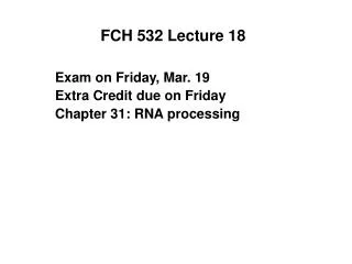
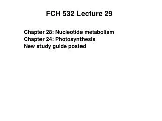
DNA replication in E. coli initiated at oriC • oriC - Unique 245-bp segment that is highly conserved in gram-negative bacteria-supports bidirectional replication. • DnaAprotein-467 aa, recognizes and binds oriC’s DnaA boxes (highly conserved 9 bp sequences) • (5’-TTATCCACA-3’) • Forms negative supercoiled DNA wrapped around 5 DnaA proteins. • Facilitated by HU and integration host factor (IHF) that cause bending of DNA. • DnaA melts 3-tandemly repeated 13 bp AT-rich segments, 5’-GATCTNTTNTTTT-3’ located near the left boundry. • Established by P1 nuclease • DnaA recruits 2 DnaB6-DnaC6 complexes to form prepriming complex. • SSB and gyrase, DnaB helixas further unwinds the DNA in prepriming complex in both directions • oriC sequence similar to RNA promoters (AT-rich) and RNAP activates primase for production RNA primers
Figure 30-29 A model for DNA replication initiation at oriC. DnaA proteins bind to DnaA boxes at oriC aided by HU and IHF 3 AT-rich 13-bp repeats are melted in ATP dependent fashion-open complex 2 DnaB6-DnaC6 complexes recruited to opposite ends of complex + 5 DnaA to form dimers. Open complex further unwound by DnaB Page 1156
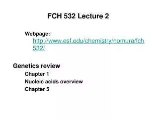
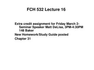
Regulation of initiation of DNA replication The doubling time of E. coli at 37 °C varies from <20 min to 10 h. Replication fork has a constant 1000 nt/s rate, meaning that the 4.6 X 10⁶ bp E. coli chromosome has a replication time, C of ~40 min. The segregationof cellular components and formation of septum has a fixed time of D = 20 min, after the completion of the corresponding round of chromosome replication. Cells with doubling times < C + D = 60 min must initiate chromosome replication before the end of the preceding cell division cycle. This results in multiforked chromosomes.
Figure 30-30 Multiforked chromosomes in E. coli. In cells that are dividing every 35 min, the fixed 60-min interval between the initiation of replication and cell division results in the production of multiforked chromosomes. Page 1157
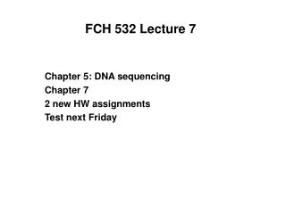
How does DNA move to the new cell? • Dam methyltransferase methylates GATC sequences in E. coli. • GATC occurs 11 times in oriC. • Newly replicated GATC segments are hemimethylated (new strands not methylated). • Hemimethylated DNA associated with the cell membrane. • Interacts with SeqA protein for the sequestration of DNA based on the oriC site.
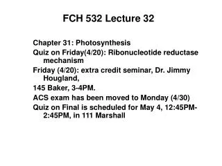
Figure 30-31 Electron micrograph of an intact and supercoiled E. coli chromosome attached to two fragments of the cell membrane. Page 1157
DNA Damage and Repair • DNA is the instruction manual for the cell. • Changes in DNA base sequence are called mutations. • Most mutations are harmful, even lethal to cells. • Silent mutations are changes in DNA sequence that do not affect the function of protein products. • Mutations can occur by spontaneous processes or induced processes. • Final error rate in E. coli replication: ~1 x 10¹⁰ base pairs. • Actual error rate of base incorporation during E. coli replication: 1 in 104-105 bases inserted. • Conclusion: repair systems correct most mismatched bases.
DNA Damage and Repair • Mutations are usually bad, but may be responsible for selective advantages, evolutionary processes. • Mutations can occur by spontaneous processes or induced processes. • Two types of spontaneous mutation processes: • Mistakes in the incorporation of deoxyribonucleotides (mismatched base pairs: A-C, G-T). • Base modifications caused by hydrolytic reactions - removal of purine base ring by hydrolysis at the N-glycosidic bond. Three types of replication errors: • Point mutation - substitution of one base pair for another. (Most common, results from base tautomerism) • Insertion of one or more extra base pairs. • Deletion of one or more base pairs.
Figure 30-51 Types and sites of chemical damage to which DNA is normally susceptible in vivo. Red, oxidation; blue, hydrolysis; green, methylation. Page 1173
Mutagenic damage to DNA caused by spontaneous processes Note conversion of cytosine to uracil in (b).
Secondary or indirect damage to DNA caused by the hydroxyl radical, OH. The OH radical is formed by interaction of The OH removes a hydrogen atom from DNA, forming H2O and a reactive DNA radical. This results in a broken DNA strand.
Some sources of ionizing radiation: Cosmic rays Medical X-rays Nuclear weapons testing (fallout) Nuclear power plants Airplane travel (high altitude) Radon gas in poorly ventilated residential buildings Radium mining tailings (waste rock - radon gas) Forgotten radium processing sites
Chemical Mutagens. • Heterocyclic base analogs like these are incorporated into replicating DNA and induce mutations by altering base-pairing characteristics.
Intercalating agents as chemical mutagens Flat , hydrophobic , typically aromatic molecules that insert between base pairs in DNA.
Figure 11.17 Binding of intercalating agents to DNA, which causes structural distortions
DNA Damage and Repair • Different responses to damage • Direct reversal of damage • Example: photolyase corrects thymine dimers • Cyclobutylthymine dimers form under UV radiation. • These pyrimidine dimers distort the DNA base pair structure. • Photolyases found in prokaryotes and eukaryotes • Bind to pyrimidine dimers and have a noncovalently bound chromophore (MTHF) that abs. 300-500nm light and transfers energy to FADH which cleaves the dimer. • Mechanism through base flipping (distortion of the double helix).
Figure 30-52 The cyclobutylthymine dimer that forms on UV irradiation of two adjacent thymine residues on a DNA strand. Page 1173
Photolyases repair pyrimidine dimers • Pyrimidine dimers distort DNA structure so that it cannot be transcribed or replicated. • A single thymine dimer is enough to kill E. coli if unrepaired. • Repaired by photolyases-bind in the dark, active in light • Use a noncovalenly bound chromophore (N5,N10-methylenyltetrahydrofolate (MTHF) or 5-deazaflavin) • absorbs 300 to 500 nm light and transfers energy to FADH- which transfers energy to break dimer. • Resulting pyrimidine anion reduces FADH• and repaired DNA is released.
Figure 30-53 X-Ray structure of E. coli DNA photolyase showing its putative DNA binding surface. Pyrimidine binding site Page 1174
DNA Damage and Repair • Alkyltransferases dealkylate alkylated nucleotides • Exposure of DNA to N-methyl-N’-nitrosoguanidine (MNNG) will alkylate purines. Reactive methylating agents shown here can convert guanine (pairs with C) to O6-methylguanine, which pairs with thymine.
Figure 30-54a The structure of E. coli Ada protein. (a) The X-ray structure of Ada’s 178-residue C-terminal segment, which contains its O6-alkylguanine–DNA alkyltransferase function. Alkyltransferases dealkylate alkylated nucleotides Transfers the alkyl group to an active Cys residue at 321. Must undergo conformational change in order to effect methyl transfer. N terminus repairs methyl phosphotriesters by binding to Cys 69 Page 1175
Figure 30-54b The structure of E. coli Ada protein. (b) The NMR structure of Ada’s 92-residue, N-terminal segment, which mediates its methyl phosphotriester repair function. Page 1175
Ada has two independent functions • C-terminus repairs O6-alkylguanine DNA through transfer of methyl group to Cys 321. • N-terminal segment repairs methyl phosphotriesters in DNA by transferring methyl group to Cys 69 • N-terminus has a Zn atom that stabilized the thiolate form over thiol form (-S- vs -SH) • -S- can attack the methyl group on DNA
Excision Repair • Two types • (1) nucleotide excision repair (NER) repairs bulky DNA lesions • (2) base excision repair (BER) repairs a single base. • NER in prokaryotes uses three subunits, eukaryotes 16 subunits. • UvrA, UvrB, and UvrC cleave the damaged DNA strand at the 7th and 3rd or 4th phosphodiester bonds from the lesion’s 5’ and 3’ sides. • Excised 11 or 12 nt oligo is displaced by UvrD.
Figure 30-55 The mechanism of nucleotide excision repair (NER) of pyrimidine photodimers. Page 1176
Excision Repair • (2) base excision repair (BER) repairs a single base. • Adenine and cytosine spontaneously deaminate to yield hypoxanthine and uracil • S-Adenosylmethionine (SAM) occasionally methylates a base to form 3-methyladenine and 7-mehtylguanine. • DNA glycosylaes cleave the glycosidic bond of altered nucleotides leaving apurinic or apyrimidinic (AP) sites. • The deoxyribose residue is cleaved on one side by the AP endonuclease, the other side by an exonuclease (DNA polymerase) and gap is filled by polymerase and DNA ligase.
Figure 30-56 Action of DNA glycosylases. These enzymes hydrolyze the glycosidic bond of their corresponding altered base (red) to yield an AP site. Page 1177
Mismatch Repair • Repairs mispairings in DNA that have not been caught by DNA polymerases and MMR can correct insertions or deletions up to 4 nt. • Must distinguish the parental strand from the daughter strand. In E. coli this is possible because the newly replicated GATC palindromes remain hemi-methylated until the Dam methyltransferase has had sufficient time to methylate the daughter strand. • Requires three proteins: MutS, MutL and MutH • MutS binds to mismatched base pair or unpaired bases as a homodimer. • The MutS-DNA complex binds to MutL homodimer • MutS-MutL translocates along the DNA forming a loop in the DNA. • Encountering a hemimethylated GATC palindrome, recruits MutH and activates single strand endonuclease to make a nick on the 5’ side of the unmethylated GATC.
Mismatch Repair • Repairs mispairings in DNA that have not been caught by DNA polymerases and MMR can correct insertions or deletions up to 4 nt. • Must distinguish the parental strand from the daughter strand. In E. coli this is possible because the newly replicated GATC palindromes remain hemi-methylated until the Dam methyltransferase has had sufficient time to methylate the daughter strand. • Requires three proteins: MutS, MutL and MutH
Mismatch Repair MutS binds to mismatched base pair or unpaired bases as a homodimer. The MutS-DNA complex binds to MutL homodimer MutS-MutL translocates along the DNA forming a loop in the DNA. Encountering a hemimethylated GATC palindrome, recruits MutH and activates single strand endonuclease to make a nick on the 5’ side of the unmethylated GATC. May be on either side of the mismatch and over 1000 bp away from it. MutS-MutL recruits UvrD helicase, which in concert with an exonuclease separates the strands and degrades the nick strand to beyond the mismatch. Gap is filled by Pol III and sealed by DNA ligase. Eukaryotes more complex; 6 homologs of MutS and 5 of MutL. MutH is exclusive to gram-negative bacteria.
Figure 30-58 The mechanism of mismatch repair in E. coli. Page 1179
SOS response • SOS response causes cells to stop dividing and repair damaged DNA. • LexA and RecA mutants always have the SOS response on. • When E. coli is exposed to agents that damage DNA, RecA mediates proteolytic cleavage of LexA. This is induced by RecA binding to ssDNA. • LexA is a repressor of 43 genes involved in DNA repair (all proceeded by 20 nt sequence called the SOS box).
Figure 30-59 Regulation of the SOS response in E. coli. Page 1180