Download
1 / 25
260 likes | 315 Views
Learn about the genome structure of vancomycin-resistant E. faecalis and its implications. Explore the mechanisms of protein expression through proteomic techniques like 2D gel electrophoresis and mass spectrometry. Unravel the role of Phages, transposons, and PTS systems in antibiotic resistance and sugar transport.

E N D
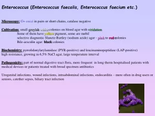
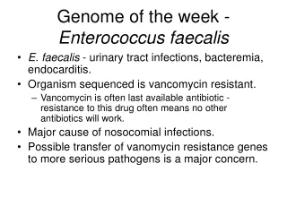
Genome of the week - Enterococcus faecalis • E. faecalis - urinary tract infections, bacteremia, endocarditis. • Organism sequenced is vancomycin resistant. • Vancomycin is often last available antibiotic - resistance to this drug often means no other antibiotics will work. • Major cause of nosocomial infections. • Possible transfer of vanomycin resistance genes to more serious pathogens is a major concern.
Genome of the week - Enterococcus faecalis • Over 25% of the E. faecalis genome consists of foreign DNA. • Phages, insertions sequences, transposons. • Likely contributed to the acquisition of resistance to multiple antibiotics. • Over 35 PTS systems • Responsible for transporting sugars into the cell. • Most found in any sequenced genome, likely utilize undigested sugars in the intestine.
Genomics DNA (Gene) Transcription Transcriptomics RNA Translation Functional Genomics PROTEIN Proteomics Enzymatic reaction METABOLITE Metabolomics The “omics” nomenclature…
Inactive mRNA RNA Degradation control Primary RNA transcript mRNA mRNA DNA Translation control RNA Transport control Modified protein protein RNA Processing control Transcriptional control Post-translational control Why study protein expression?
2D gel electrophoresis • Method for separating and visualizing proteins • Separation by charge and mass • Mass spectrometry • High throughput analysis and identification of proteins. • Fragmentation of proteins • Analysis of peptides • Book - pages 273-300.
2D-SDS PAGE gel The first dimension (separation by isoelectric focusing) - gel with an immobilised pH gradient - electric current causes charged proteins to move until it reaches the isoelectric point (pH gradient makes the net charge 0) What determines the charge of a protein?
4 5 Stable pH gradient 6 7 8 9 10 Isoelectric point (pI) • Separation by charge: Low pH: Protein is positively charged At the isolectric point the protein has no net charge and therefore no longer migrates in the electric field. High pH: protein is negatively charged
2D-SDS PAGE gel The first dimension (separation by isoelectric focusing) - gel with an immobilised pH gradient - electric current causes charged proteins to move until it reaches the isoelectric point (pH gradient makes the net charge 0) The second dimension (separation by mass) -pH gel strip is loaded onto a SDS gel -SDS denatures the protein (to make movement solely dependent on mass, not shape) and eliminates charge.
Advantages vs. Disadvantages • Not for hydrophobic proteins • Limited by pH range • Not easy for low abundant proteins • Analysis and quantification are difficult • Good resolution of proteins • Detection of posttranslational modifications
Current Mass Spec Technologies • Proteome profiling/separation • 2D SDS PAGE - identify proteins • 2-D LC/LC - high throughput analysis of lysates (LC = Liquid Chromatography) • 2-D LC/MS (MS= Mass spectrometry) • Protein identification • Peptide mass fingerprint • Tandem Mass Spectrometry (MS/MS) • Quantative proteomics • ICAT (isotope-coded affinity tag) • ITRAQ
Mass Spectrometry (MS) • Introduce sample to the instrument • Generate ions in the gas phase • Separate ions on the basis of differences in m/z with a mass analyzer • Detect ions
2D - LC/LC Peptides all bind to cation exchange column (trypsin) Study protein complexes without gel electrophoresis Successive elution with increasing salt gradients separates peptides by charge Peptides are separated by hydrophobicity on reverse phase column Complex mixture is simplified prior to MS/MS by 2D LC
Identifying proteins • Trypsin - digest your protein • Digests after R and K amino acids. • Run peptide fragments on mass spec • Digest the protein database “in silico” • Compare mass spec data with theoretical data. • What must be true to identify your protein?
Artificially trypsinated Fragmented using trypsin Artificial spectra built Spot removed from gel Protein Identification by MS Spectrum of fragments generated MATCH Library Database of sequences (i.e. SwissProt)
How protein sequencing works Ser-Glu-Leu-Ile-Arg-Trp • Use Tandem MS: two mass analyzer in series with a collision cell in between • Collision cell: a region where the ions collide with a gas (He, Ne, Ar) resulting in fragmentation of the ion • Fragmentation of the peptides in the collision cell occur in a predictable fashion, mainly at the peptide bonds • The resulting daughter ions have masses that are consistent with known molecular weights of dipeptides, tripeptides, tetrapeptides… Collision Cell Ser-Glu-Leu-Ile-Arg Ser-Glu-Leu-Ile Ser-Glu-Leu Etc…
Advantages vs. Disadvantages • High capital costs • Requires sequence databases for analysis • Determination of MW and aa. Sequence • Detection of posttranslational modifications • High-throughput capability
ISOTOPE-CODED AFFINITY TAG (ICAT): a quantitative method • Label protein samples with heavy and light reagent • Reagent contains affinity tag and heavy or light isotopes Chemically reactive group: forms a covalent bond to the protein or peptide Isotope-labeled linker: heavy or light, depending on which isotope is used Affinity tag: enables the protein or peptide bearing an ICAT to be isolated by affinity chromatography in a single step
Example of an ICAT Reagent Biotin Affinity tag: Binds tightly to streptavidin-agarose resin Reactive group: Thiol-reactive group will bind to Cys Linker: Heavy version will have deuteriums at * Light version will have hydrogens at *
100 0 0 600 200 400 550 570 590 How ICAT works? Affinity isolation on streptavidin beads Lyse & Label Quantification MS Identification MS/MS NH2-EACDPLR-COOH Light 100 MIX Heavy Proteolysis (eg trypsin) m/z m/z
Advantages vs. Disadvantages • Yield and non specificity • Slight chromatography differences • Expensive • Tag fragmentation • Meaning of relative quantification information • No presence of cysteine residues or not accessible by ICAT reagent • Estimates relative protein levels between samples with a reasonable level of accuracy (within 10%) • Can be used on complex mixtures of proteins • Cys-specific label reduces sample complexity • Peptides can be sequenced directly if tandem MS-MS is used


![Trivedi Effect - Impact of an external energy on Enterococcus faecalis [ATCC – 51299] in relation to antibiotic suscepti](https://cdn4.slideserve.com/7685708/5-14-2015-dt.jpg)