Download
1 / 23
240 likes | 397 Views
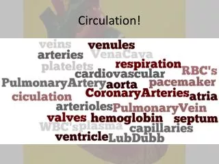
Chapters 32 and 33. Circulation. Circulation and Circulatory Systems heart atria : receive blood ventricles : pump blood veins vessels that transport blood back to heart all veins carry deoxygenated blood (CO 2 -rich) , except those leaving the lungs

E N D
Chapters 32 and 33 Circulation
Circulation and Circulatory Systems • heart • atria: receive blood • ventricles: pump blood • veins • vessels that transport blood back to heart • all veins carry deoxygenated blood (CO2-rich) , except those leaving the lungs • connective tissue thin layer of smooth muscle elastin epithelial tissue • contain one-way valves prevent backflow • venules • smallest veins in body leave capillaries join larger veins • arteries • vessels that transport blood away from heart • all arteries carry oxygenated blood (O2-rich) , except those bringing blood to lungs • connective tissue thick layer of smooth muscle elastin epithelial tissue • arterioles • smallest arteries in body merge into capillaries • capillaries • smallest blood vessels in body • thin layer of epithelial tissue • gas and nutrient exchange • capillary beds
blockages can be a problem in all vessels • especially arteries Fig. 32.3 Arteries, capillaries, and veins
Some Animals Lack a Circulatory System • utilize diffusion, osmosis, or cell-to-cell transport • Invertebrate Circulatory Systems • open circulatory systems • blood enters and then leaves the vessels • after leaving, blood fills hemocoels (“blood cavities”) • saturates body tissues in blood muscular contractions blood returns to heart • closed circulatory systems • more efficient • blood remains in vessels • blood flows much more rapidly
Vertebrate Circulatory Systems • all vertebrates have closed systems • blood • a connective tissue; 0.9% saline • formed elements (cell types) – 45% • erythrocytes (rbc’s) • transport O2 t0 body (hemoglobin) • concave shape and no nucleus • spleen • removes old rbc’sand stores wbc’s • leukocytes (wbc’s) • many different types function in the immune system • thrombocytes (platelets) • cellular fragments that play a major role in blood clotting • all derive from hemocytoblasts in red bone marrow • plasma (a watery substance) – 55% • 92% H2O, 7% proteins, 1% other solids, gases, wastes • plasma proteins • albumins, globulins, fibrinogen, prothrombin, etc. • salts, fats, glucose, amino acids, hormones, vitamins, ions, etc. • gases: extra O2, CO2 • nitrogenous wastes: urea, uric acid Fig. 31.4 Formed elements
blood clotting • 7-10 steps; about 15 substances involved • some substances prevent accidental clotting • three major reactions • platelets form a “plug” at wound site release thromboplastin • thromboplastin and Ca+ ions convert prothrombin to thrombin • thrombin converts fibrinogen to fibrin fibers seal wound • the damaged cells, collagen, and other substances also help seal wound Fig. 32.14 Blood clotting
fish • two-chambered heart • 1 atrium, 1 ventricle • gas exchange across gill capillaries • amphibians and most reptiles • three-chambered heart • 2 atria, 1 ventricle • systemic vs. pulmonary circuit • pulmonary: blood flow to and from lungs • systemic: blood flow to and from rest of body • the two are not completely separate • deoxygenated vs. oxygenated blood • some mixing occurs in the single ventricle • birds, crocodilians, mammals, humans • four-chambered heart • 2 atria, 2 ventricles • systemic and pulmonary circuits completely separate • normally no mixing of deoxygenated and oxygenated blood
Fig. 32.5 Comparison of circulatory systems in vertebrates
Human Circulatory System • path of blood flow through body • deoxy. blood in body capillaries body venules body veins superior/inferior vena cavas right atrium right AV valve right ventricle pulmonary semilunar valve pulmonary artery lung arterioles lung capillaries (gas exchange; blood now oxy.) lung venules pulmonary veins left atrium left AV valve left ventricle aortic semilunar valve aorta body arteries body arterioles body capillary beds (gas & nutrient exch.; blood now deoxy.) REPEAT • blood makes a complete circuit with every beat
Fig. 32.7 Internal view of the heart Fig. 32.8 Heart valves
control of heart contractions • unique nature of heart (cardiac) muscle • branching of muscle fibers • extrinsic control • external control outside of heart • nervous system and hormones (esp., epinephrine) • speeds up or slows down heart rate • intrinsic control • control within heart itself • sinoatrial (SA) and atrioventricular (AV) nodes • specialized regions of cells that can generate and carry electrical impulses • ventricular septum and Purkinje fibers • SA node creates impulse walls of atria atria contract signal travels to AV node signal routed down ventricular septum in two paths Purkinje fibers walls of ventricles ventricles contract • SA node initiates heartbeat; AV signals ventricles to contract
the working heart • heart beat and activity • 1st sound: right and left AV valves closing • 2nd sound: aortic and pulmonary semilunar valves closing • all valves prevent backflow of blood • can be monitored with an EKG (electrocardiogram) • systole vs. diastole • stroke volume vs. cardiac volume • heart rate (pulse) – average = 72 beats/minute • blood pressure • sphygmomanometer • systolic vs. diastolic pressure (mm Hg) • normal measurements (120/80) • arterioles • vasoconstriction muscle walls thicken increases blood pressure • vasodilation muscle walls thin decreases blood pressure • all gas and nutrient exchange takes place across capillaries • blood pressure lowest in veins (venous blood) • movement assisted by valves and smooth/skeletal muscle contraction
human circulatory circuits • circuit: a major pathway of blood flow and return • pulmonary • systemic • hepatic portal • nutrients absorbed by small intestine travel in hepatic portal vein to liver liver monitors blood content and stores extra nutrients blood enters general circulation • renal • cardiac • coronary arteries and veins • numerous ones in head and brain
Lymphatic System • series of small vessels that parallel circulatory system • transports lymph instead of blood • colorless, interstitial fluid that is derived from tissues • may be in tissue cells or between tissue layers • may be blood plasma that seeps into tissues • fluid moves in same fashion as venous blood merges with circ. system • four essential functions • maintain fluid and ion balances in body • transports certain fatty acids • part of the immune system and cooperates with it • route by which interstitial fluids can return to the circ. system • structures • lymph capillaries larger lymph vessels • lymph nodes • mass of lymphoid tissue located along the course of a lymph vessel • highly involved with immune system • lymph organs • other organs strongly associated with lymph • spleen, bone marrow, tonsils, thymus gland, etc.