Download
1 / 30
300 likes | 413 Views
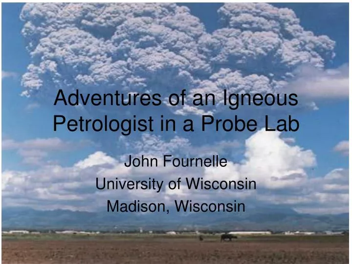
Adventures of an Igneous Petrologist in a Probe Lab. John Fournelle University of Wisconsin Madison, Wisconsin. Mt. Pinatubo June 15, 1991: 20th century’s 2nd largest (volumetric) eruption largest observed SO 2 cloud, 20 megatons. Anhydrite (CaSO 4 )…in a volcanic rock??!!!.

E N D
Adventures of an Igneous Petrologist in a Probe Lab John Fournelle University of Wisconsin Madison, Wisconsin
Mt. Pinatubo • June 15, 1991: • 20th century’s 2nd largest (volumetric) eruption • largest observed SO2 cloud, 20 megatons
Anhydrite (CaSO4)…in a volcanic rock??!!! • Anhydrite had been observed in the pumices from the SO2-rich eruption of El Chichon in 1982 • Experiments had proven that it truly was an equilibrium phase in a hydrous oxidized dacitic magma • It is rarely observed in pumices which have been rained upon/weathered • When I learned of the SO2-rich Pinatubo eruption, I guessed that anhydrite might be present
Optically, high birefringence (~like calcite) Above 3 BSE images: anhydrite as microphenos (with apatite!); and as inclusions in plag and hb BSE: brighter than plag, darker than apatite; Reflected light: distinctive surface (rough, ~soft/scratches?)
Plan: A student to do single crystal XRD…to confirm same crystal structure (there are several forms between gypsum and anhydrite -- First separate CaSO4 from Ca5(PO4)3 grains by EDS (heavy liquid separation from pumice)-- But we found “bumps” all over the CaSO4 when we looked at SEM images with the electron microprobe!
Project Redefined: Are the pyramids laboratory artifacts? Are they produced during eruption? Are they produced before the eruption, at depth? Is the anhydrite “true” high temperature orthorhombic anhydrite? What are the pyramids? What caused them?
Pyramids formed before glass vesciculated, so not laboratory artifact -- and also before eruption
Single crystal XRD: Yes, phenocrysts true high temperature orthorhombic anhydrite
Anhydrite -- trapped inside large plagioclase…anhydrite has pyramids that had formed prior to capture… evidence that pyramids formed at depth
Electron Backscatter Diffraction: both pyramid and host anhydrite inclusion in plagioclase are orthorhombic CaSO4 1=Pyramid 2=Host
Comparison: GaN grown in laboratory vs Pinatubo magma chamber
Conclusion • Textures suggest that anhydrite pyramids grew from vapor deposition in the magma chamber prior to eruption • This is consistent with temperatures, SO2 content of eruptive gas, magma fO2 and water contents • This also is consistent with the evidence for “excess” sulfur that cannot be accounted for strictly from magma sulfur solubility; i.e. this supports the suggestion of Gerlach that there was a previously separated sulfur-rich gas below the volcano • We give this the name “magmatic vapor deposition” Jakubowski, Fournelle, Welch, Swope and Camus (2002) Evidence for magmatic vapor deposition of anhydrite prior to the 1991 climactic eruption of Mount Pinatubo, Philippines. American Mineralogist, 87, 1029-1045.
Problems of Secondary Fluoresence in EPMA Issue: Is there ~10 wt% Nb in the Pd3Hf and Pd2HfAl?
Results from other research group using EDS (Nb Ka) My results using WDS (Nb La)
Results from other research group using EDS (Nb Ka) My results using WDS (Nb La)
Nb Ka is really there in the spectrum! But it shouldn’t be, right??? Well, yes and no…
Two approaches to understanding the problem of “secondary fluorescence across phase boundaries”: • Experimental: create a “non-diffused couple”, putting two perfectly flat pieces of A and B together …but not always easy nor possible • Simulate with a model that reproduces the physics of electron-material interactions and follows the x-rays; i.e. the Monte Carlo PENEPMA program (based upon PENELOPE)
PENEPMA simulation of secondary fluorescence: 5 microns away from the Nb, inside the Pd2HfAl
Comparing experimental (non-diffused couple) with PENEPMA: geometry effects also
Al-Ir compounds and the problem of light element-heavy element matrix corrections in EPMA Problem: with such a large difference in Z (13-77), using pure end member standards, there is a potential for major errors in the matrix correction. This has been noted by some researchers, but not systematically addressed before. Figure: BSE image of alloy Ir-46.20Al annealed at 1130oC for more than 1000 hours The bright phase is fcc-(Ir) and the dark one is B2-AlIr
Compounding this problem are at least two issues • MAC’s not known well: Al ka by Ir has 26% range • Heinrich (1966): 2351 • Henke (1985): 2057 • FFAST (Chandler 2005): 2057 • Mean atomic number of the intermediate alloy also controversial (important for backscatter correction)
PENEPMA Simulations for 6 intermediate compounds were run, to find which one closest fit the actual experimental EPMA data. -- Ir matches for 50, but AL matches for 53. Split the difference: 51.5 as the most probable true composition… -- Next step is to set up a geometry for secondary fluorescence to see if that would improve the fit