Download
1 / 21
220 likes | 227 Views
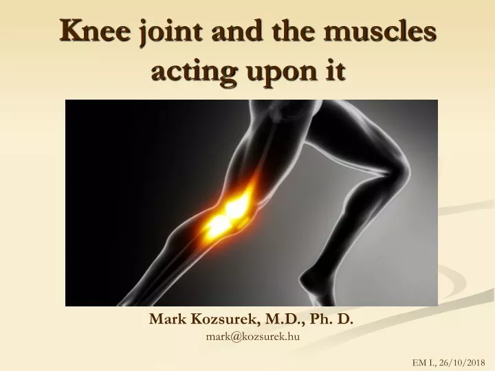
Knee joint and the muscles acting upon it. Mark Kozsurek, M.D., Ph. D. mark@kozsurek.hu. EM I., 26/10/2018. Knee is the biggest joint of the human body. I t bears enormous weight and pressure loads , so not surprisingly its frequently injured or is involved in osteoarthritis !

E N D
Kneejoint and themusclesactinguponit Mark Kozsurek, M.D., Ph. D. mark@kozsurek.hu EM I., 26/10/2018
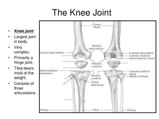
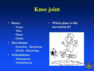
Knee is thebiggestjoint of the human body. It bears enormous weight and pressure loads, sonotsurprisinglyitsfrequentlyinjuredor is involved in osteoarthritis! • Letsdiscussthekneejoint in thefollowingorder (recommendedforallthejoints – nothingwill be missed): • articularfacets: theshape of articulatingsurfacespredictsthetype of joint and thepossiblemovements. Wehaveto mention here thetwomeniscias non-obligatoryfeatures. • jointcapsule: howdoesitsupportorrestrictmovements? • ligaments: mayincreasestabilitybutcanalso limit movements • type of joint, movements • groups of musclesactinguponthekneejoint
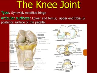
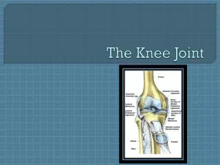
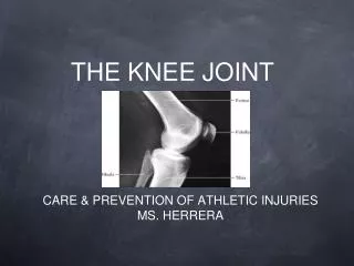
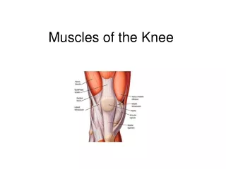
Articularfacets Knee joint is formedbythearticulation of thetwofemoralcondyles, thetwotibialcondyles and thepatella! Contribution of fibula is indirect!
Menisci Articular incongruencyexistsbetweenfemoral and tibialcondyles and forcompensationmenisciareinsertedbetweenthem. The sameforce is spreadalong a muchbiggersurfaceresulting a lowerpressurethat is welltoleratedbythehyalinecartilage attached to the capsule: injures more frequently Only the horns of menisci are attached tothe tibia, so they can glide freely between the codyles, and even can form wider or narrower demilunes!
Articular capsule of theknee fibrous membrane fibrocyte nuclei synovial membrane
synovial membrane fibrous membrane Note that anteriorly and posteriorly synovial and fibrous layers of the capsule are getting divorced and the gap is filled by fat pads. Intraarticular (cruciate) ligaments are invested by the synovial membrane.
synovial membrane fibrous membrane Many bursae can be found close to the knee joint: in general they reduce friction. Suprapatellar bursa may exit separately, but may also merge with the suprapatellar recess. Suprapatellar recess might be considered as a reserve fold: without that the anterior portion of the joint capsule would arrest movement during flexion too early. (Normally flexion is only limited by the amount of soft tissues – fat and/or muscle.) Suprapatellar bursa Suprapatellar recess Prepatellar bursa Superficial and deep infrapatellar bursa
Articular ligaments Lateralcollateral ligament descendsfromthecondyle of thefemurtothehead of fibula as a roundedstructure and is completlyindependentfromthecapsule. Medialcollateral ligament is flat and is attachedtightlytothecapsule and themedialmeniscus. Collateralligamentsstabilizethejointonthetwosides and inhibithyperextension. Theyaretense in extendedposition and relaxwhentheknee is flexed. extraarticular vs. intraarticular ligaments
Cruciate ligamentsrestrictdisplacement of thetibial and femoralcondyli and supportrollingmovements of articulatingsurfacesinstead of gliding. Cruciate ligamentsaremuch more important in theflexedposition of theknee, whencollateralligamentsareloose! drawer test
Tendons There are several tendons inserting around the knee: the most of them merge with the capsule and reinforce it. PES ANSERINUS Flattened tendon of Quadriceps femoris muscle inserts at the base of patella, then continues downward from the apex of patella to the tibial tuberosity as the patellar ligament. Thickenned lateral portion of the fascia lata terminates at the lateral condyle of tibia as the iliotibial tract. On the medial condyle Sartorius, Gracilis and Semitendinosus muscles insert by forming the (superficial) pes anserinus. (Deep pes anserinus is the branching pattern of the tendon of Semimembranosus muscle.)
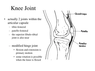
Mechanism of movement Forthefirstglance, kneeseemsto be a hingejointwithonetransverseaxisaroundwhichflexion and extension is available. Butwhenit is flexed, collateralligamentsrelax and a newaxiswithlongitudinalorientationappearsmakinginward and outwardrotationavailable. MODIFIED HINGE JOINT: uniaxialwhenextended, biaxial in flexedposition
FullextensionperformedbyQudricepsfemoris „locks” theknee (this is alsocalled „screw-homemechanism”) reducing the amount of muscle work needed to maintain the standing position. During the last 30 degrees of knee extension, the tibia (open chain) must rotateexternally or femur (closed chain) internally about 10 degrees. In thispositionthe broad and flat areas on the inferior aspects of thefemoralcondyles rest onthetibialplateaus, alltheligamentsarestretched and thejoint is stable! Atthebeginning of flexion, Politeusmusclerotatestibiamedially (openchain) and/orfemurlaterally (closedchain), the curved and rounded areas of femoral condylesget in contactwiththetibialcodyles, ligamentsrelax and thejoint is unlocked. Furtherflexion is performedbythehamstringmusclesmainly.
Musclesmovingthekneejoint EXTENSORS FLEXORS
ADDUCTORS Little effectuponthekneejoint, theymainlyact in thehipjoint!
Knee joint ismainly moved by the muscles of the thigh. Anteriorly the extensor compartment containing the Quadriceps femoris and Sartorius muscles is found. (Intrestingly, Sartorius muscle is in the extensor compartment but acts as a flexor upon the knee joint!) Posteriorly the flexor group constituted by the Biceps femoris, Semitendinosus and Semimembranosus muscles is seen. These act not only as flexors, but in the flexed position, when collateral ligaments are loose, also rotate the leg medially (Semitendinosus and Semimembranosus) or laterally (Biceps femoris). Adductors found medially rather act upon the hip joint and are not discussed here! EXTENSORS ADDUCTORS FLEXORS
Popliteal fossa Rhomboid area bounded by the thigh flexor tendons (medially: Semitendinosus and Semimembranosus, laterally: Biceps femoris) superiorly, and by the medial and lateral bellies of the Gastrocnemius inferiorly. Base is constituted by the knee joint capsule and the Popliteus muscle. Prominent structures of the region are: popliteal artery and vein, small saphenous vein, tibial and common peroneal nerves. Some lymph nodes can also be found here embedded into the adipous tissue of the fossa.
Mosaicplasty (OATS: osteochondralautografttransfersystem)