Download
1 / 7
80 likes | 504 Views
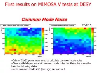
Common B-MODE ARTIFACTS. Anisotropy Acoustic shadowing Reverberation Refractile shadowing Enhanced through-transmission. ANISOTROPY: Transducer not perpendicular. Biceps tendon bright. Biceps tendon dark. Humerus. Humerus. Transverse.

E N D
Common B-MODE ARTIFACTS • Anisotropy • Acoustic shadowing • Reverberation • Refractile shadowing • Enhanced through-transmission
ANISOTROPY: Transducer not perpendicular Biceps tendon bright Biceps tendon dark Humerus Humerus Transverse Anisotropy is a sonographicartifact, especially relevant in tendons that occurs when the ultrasound beam does not insonate perpendicular to the tendon
ANISOTROPY: Object not perpendicular Toe end Heel end Biceps tendon bright Biceps tendon dark Humerus Humerus Longitudinal The probe should be maintained parallel to thetendon. In theeventthattheobject (region of interest) isnot perpendicular tothetransducer , non uniformpressure can beapplied –in this case, pressing downslightlyharder at theheelendwillensurethatthetendonis perpendicular.
ACOUSTIC SHADOWING Calcification S Acoustic shadowing below a dense calcification. As no significant sound is passing underneath the calcification and returning to the transducer, a black ‘shadow’ (S) is observed.
REVERBERATION (Comet tail or ring down artifact) Reverberation This occurs when there are two very reflective surfaces which are closing spaced as are seen in needles or other metal work. This imae shows a knee being aspirated from the right side. A reverberation artifact is seen below the needle.
REFRACTILE SHADOWING T This is particularly seen with tendons (T). It occurs because the US beam is hitting a structure of a different acoustic impedance at an oblique angle. In this case, the hypo-echoic areas (arrows) should not be mistaken for tenosynovitis.
ENHANCED THROUGH-TRANSMISSION C This occurs when there is over-compensation of returning echoes as the sound waves have passed through the liquid filled structure e.g. cyst (C) more quickly than expected. As a result, the area below appears more echogenic (arrows) than usual.