Download
1 / 15
150 likes | 167 Views
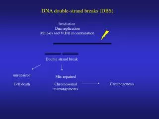
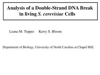
Analysis of a Double-Strand DNA Break in living S. cerevisiae Cells. Leana M. Topper*, L. Kevin Lewis # , Kerry S. Bloom*, and Michael A. Resnick #. *Department of Biology, University of North Carolina at Chapel Hill; # National Institute of Environmental Health Sciences,

E N D
Analysis of a Double-Strand DNA Break in living S. cerevisiae Cells Leana M. Topper*, L. Kevin Lewis#, Kerry S. Bloom*, and Michael A. Resnick# *Department of Biology, University of North Carolina at Chapel Hill; #National Institute of Environmental Health Sciences, NIH Research Triangle Park
Chromosome III MAT CEN ~85 kb LacO array ~500 bp
MAT LacO KpnI KpnI probe 2.5 kb No pGalHOT W/ pGalHOT 0 0.5 2 3 4 1 0 0.5 2 3 4 1 h post gal 8 kb KpnI fragment HO cut fragment
Live cell after HO induction 6 min 7 min 2 min 13 min 8 min 10 min 15 min
Movement of Spindle Pole Bodies and lacO in live cells following HO expression Average film time: 13 min No bud: 13 Small-budded cells: 8 Large-budded cells: 29 Average spindle length = 1.69 ±0.32mm Range = 1.05-2.44 mm
Deletion of Rad52 does not affect LacO movement 3 min 6 min 0 min 11 min 13 min 18 min
Rad52-GFP foci and Spindle Pole Bodies Move Independently 1 min 5 min 7 min 17 min 8 min 12 min 18 min