Download
1 / 56
560 likes | 571 Views
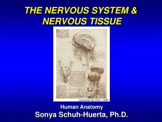
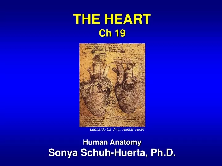
THE HEART Ch 19 Human Anatomy Sonya Schuh-Huerta, Ph.D. Leonardo Da Vinci, Human Heart. The Heart: An Amazing Piece of Machinery. Although it’s not where you keep your emotions, it’s an amazing vital organ of your body.

E N D
THE HEART Ch 19 Human Anatomy Sonya Schuh-Huerta, Ph.D. Leonardo Da Vinci, Human Heart
The Heart: An Amazing Piece ofMachinery • Although it’s not where you keep your emotions, it’s an amazing vital organ of your body
The Heart • A muscular double pump • Pulmonary circuit takes blood to & from the lungs • Systemic circuit vessels transport blood to & from body tissues • Atria receive blood from the pulmonary & systemic circuits • Ventricles the muscular pumping chambers of the heart
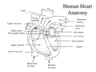
The Pulmonary & Systemic Circuits Capillary beds of lungs where gas exchange occurs Pulmonary Circuit Pulmonary arteries Pulmonary veins Aorta and branches Venae cavae Left atrium Right atrium Left ventricle Heart Right ventricle Systemic Circuit Capillary beds of all body tissues where gas exchange occurs Oxygen-rich, CO2-poor blood Oxygen-poor, CO2-rich blood
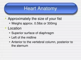
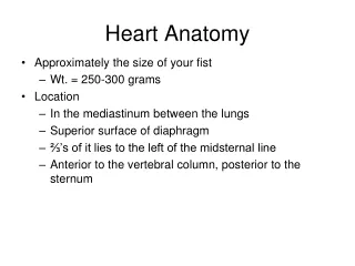
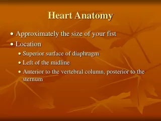
Location & Orientation Within the Thorax • Heart typically weighs 250–350 grams (~healthy heart) • Largest organ of the mediastinum • Located between the lungs • Apex lies to the left of the midline • Base is the broad posterior surface
Midsternal line Rib 2 Diaphragm (a) Mediastinum Heart Right lung Posterior (b) Location of the Heart in the Thorax Superior vena cava Aorta Parietal pleura (cut) Pulmonary trunk Left lung Pericardium (cut) Apex of heart Diaphragm (c) Mediastinum Superior vena cava Aorta Left lung Right auricle of right atrium Fat in epicardium Rib 5 Right ventricle Pericardium (cut) Apex of heart (d)
Structure of the Heart – Coverings • Pericardium – 2 primary layers • Fibrouspericardium • Strong layer of dense connective tissue • Serous pericardium • Formed from 2 layers • Parietal layer of the serous pericardium • Visceral layer of the serous pericardium (= epicardium)
Layers of the Pericardium & of the Heart Wall Pericardium Myocardium Pulmonary trunk Fibrous pericardium Parietal layer of serous pericardium Pericardial cavity Epicardium (visceral layer of serous pericardium) Heart wall Myocardium Endocardium Heart chamber
Structure of the Heart – Layers of the Heart Wall • Epicardium • Visceral layer of the serous pericardium • Myocardium • Consists of cardiac muscle – all the muscle of the heart • Muscle arranged in circular & spiral patterns • Endocardium • Endothelium resting on a layer of connective tissue • Lines the internal walls of the heart
Cardiac muscle bundles Circular & Spiral Arrangements of Cardiac Muscle Bundles
Heart Chambers • Right & left atria • Superior chambers • Right & left ventricles • Inferior chambers • Internal divisions • Interventricular septa • Interatrial septa • External markings • Coronary sulcus • Anterior interventricular sulcus • Posterior interventricular sulcus
Gross Anatomy of the Heart Left common carotid artery Brachiocephalic trunk Left subclavian artery Aortic arch Superior vena cava Ligamentum arteriosum Right pulmonary artery Left pulmonary artery Ascending aorta Left pulmonary veins Pulmonary trunk Auricle of left atrium Right pulmonary veins Circumflex artery Right atrium Left coronary artery (in coronary sulcus) Right coronary artery (in coronary sulcus) Anterior cardiac vein Left ventricle Right ventricle Great cardiac vein Right marginal artery Anterior interventricular artery (in anterior interventricular sulcus) Small cardiac vein Inferior vena cava Apex
Inferior View of the Heart Aorta Superior vena cava Right pulmonary artery Left pulmonary artery Right pulmonary veins Left pulmonary veins Right atrium Auricle of left atrium Left atrium Inferior vena cava Great cardiac vein Coronary sinus Right coronary artery (in coronary sulcus) Posterior vein of left ventricle Posterior interventricular artery (in posterior interventricular sulcus) Left ventricle Middle cardiac vein Right ventricle Apex (d) Inferior view; surface shown rests on the diaphragm (base of heart).
Right Atrium • Forms right border of heart • Receives blood from systemic circuit • Pectinate muscles • Ridges inside anterior of right atrium • Crista terminalis • Landmark used to locate veins entering right atrium • Fossa ovalis • Depression in interatrial septum • Remnant of foramen ovale (from embryonic developmt)
Right Ventricle • Receives blood from right atrium through the tricuspid valve • Pumps blood into pulmonary circuit via • Pulmonary trunk • Internal walls of right ventricle • Trabeculae carneae • Papillary muscles • Chordae tendineae • Pulmonary semilunar valve • Located at opening of right ventricle & pulmonary trunk
Heart Chambers Aorta Left pulmonary artery Superior vena cava Left atrium Right pulmonary artery Left pulmonary veins Pulmonary trunk Right atrium Mitral (bicuspid) valve Right pulmonary veins Fossa ovalis Aortic valve Pectinate muscles Pulmonary (semilunar) valve Tricuspid valve Left ventricle Right ventricle Papillary muscle Chordae tendineae Interventricular septum Trabeculae carneae Epicardium Inferior vena cava Myocardium Endocardium (e) Frontal section
Left Atrium • Makes up heart’s posterior surface • Receives oxygen-rich blood from lungs through pulmonary veins • Opens into the left ventricle through: • Mitral valve (= bicuspid;left atrioventricular valve)
Left Ventricle • Forms apex of the heart • Very thick strong muscle • Internal walls of left ventricle • Trabeculae carneae • Papillary muscles • Chordae tendineae • Pumps blood to systemic circuit via • Aortic semilunar valve (= aortic valve)
Heart Valves • Each valve composed of • Endocardium with connective tissue core • Atrioventricular (AV) valves • Between atria & ventricles tricuspid & bicuspid • Aortic & pulmonary valves • At junction of ventricles & great arteries pulmonary & aortic semilunar
Heart Chambers & Valves Aorta Left pulmonary artery Superior vena cava Left atrium Right pulmonary artery Left pulmonary veins Pulmonary trunk Right atrium Mitral (bicuspid) valve Right pulmonary veins Fossa ovalis Aortic (semilunar) valve Pectinate muscles Pulmonary (semilunar) valve Tricuspid valve Left ventricle Right ventricle Papillary muscle Chordae tendineae Interventricular septum Trabeculae carneae Epicardium Inferior vena cava Myocardium Endocardium (e) Frontal section
Fibrous Skeleton • Surrounds all 4 valves • Composed of dense connective tissue • Functions: • Anchors valve cusps • Prevents overdilation of valve openings • Main point of insertion for cardiac muscle • Blocks direct spread of electrical impulses
Fibrous Skeleton & Valve Structure Pulmonary valve Aortic valve Area of cutaway Mitral valve Tricuspid valve Tricuspid (right atrioventricular) valve Mitral (bicuspid, left atrioventricular) valve Aortic valve Myocardium Pulmonary valve Fibrous skeleton Anterior
The Beating Heart Blood returning to the heart fills atria, putting pressure against atrioventricular valves; atrioventricular valves are forced open. 1 Direction of blood flow Atrium Cusp of atrioventricular valve (open) Chordae tendineae 2 As ventricles fill, atrioventricular valve flaps hang limply into ventricles. Papillary muscle Ventricle 3 Atria contract, forcing additional blood into ventricles. (a) AV valves open; atrial pressure greater than ventricular pressure
The Beating Heart 1 Ventricles contract, forcing blood against atrioventricular valve cusps. Atrium Cusps of atrioventricular valve (closed) 2 Atrioventricular valves close. Blood in ventricle 3 Papillary muscles contract and chordae tendineae tighten, preventing valve flaps from everting into atria. (b) AV valves closed; atrial pressure less than ventricular pressure
The Beating Heart Aorta Pulmonary trunk As ventricles contract and intraventricular pressure rises, blood is pushed up against semilunar valves, forcing them open. (a) Semilunar valves open As ventricles relax and intraventricular pressure falls, blood flows back from arteries, filling the cusps of semilunar valves and forcing them to close. (b) Semilunar valves closed
Heart Sounds • “Lub-dub” sound of valves closing • First sound “lub” • The AV valves closing • Second sound “dub” • The semilunar valvesclosing
Heart Sounds Pulmonary valve “Lub” Aortic valve Area of cutaway Mitral valve Myocardium Tricuspid valve Tricuspid (right atrioventricular) valve Mitral (left atrioventricular) valve Aortic valve “dub” Pulmonary valve Fibrous skeleton Anterior
Pathway of Blood Through the Heart • Beginning with oxygen-poor blood in the superior & inferior venae cavae • Go through pulmonary & systemic circuits • Blood passes through all structures sequentially • Atria contract together • Ventricles contract together
Pulmonary semilunar valve Tricuspid valve Superior vena cava (SVC) Inferior vena cava (IVC) Coronary sinus Right ventricle Pulmonary trunk Right atrium Pulmonary trunk SVC Coronary sinus Tricuspid valve Right atrium Pulmonary semilunar valve IVC Right ventricle Oxygen-poor blood returns from the body tissues back to the heart. Two pulmonary arteries carry the blood to the lungs (pulmonary circuit) to be oxygenated. To heart To lungs Pulmonary arteries Oxygen-rich blood Oxygen-poor blood Oxygen-rich blood is delivered to the body tissues (systemic circuit). Oxygen-rich blood returns to the heart via the four pulmonary veins. To body To heart Aorta Pulmonary veins Mitral valve Aortic semilunar valve Left atrium Left ventricle Aortic semilunar valve Mitral valve Four pulmonary veins Left ventricle Left atrium Aorta Blood Flow Through the Heart
Heartbeat • 70–80 beats per minute at rest (adult) • Systole contraction of a heart chamber • Diastole expansion (relaxation) of a heart chamber • Systole & diastole also refer to • Stage of heartbeat when ventricles contract & expand
Structure of Heart Wall • Walls differ in thickness • Atria thin walls • Ventricles very thick walls (especially left) • Systemic circuit • Longer than pulmonary circuit • Has greater resistance to blood flow
Structure of Heart Wall • Left ventricle:3 times thickerthan right • Exerts more pumping force • Flattens right ventricle into a crescent shape Left ventricle Right ventricle Interventricular septum
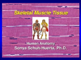
Cardiac Muscle Tissue • Forms a thick layer called myocardium • Striated like skeletal muscle • Contractions pump blood through the heart & into blood vessels
Cardiac Muscle Tissue • Cardiac muscle cells • Short • Branching • Have 1 or 2 nuclei • Not fused muscle cells like skeletal muscle fibers
Cardiac Muscle Tissue • Cells join at intercalated discs • Complex junctions • Form cellular networks • Cells are separated by delicateendomysium • Binds adjacent cardiac fibers • Contains blood vessels & nerves
Intercalated Discs • Intercalated discs complex junctions • Adjacent sarcolemmas interlock • Possess 3 types of cell junctions • Desmosomes • Fasciae adherens long desmosome-like junctions • Gap junctions
Nucleus Intercalated discs Cardiac muscle cell Gap junctions Fasciae adherens (a) Microscopic Anatomy of Cardiac Muscle Cardiac muscle cell Mitochondrion Intercalated disc Nucleus Mitochondrion T tubule Sarcoplasmic reticulum Z disc Nucleus Sarcolemma (b) I band A band I band
Cardiac Muscle Tissue • Triggered to contract by Ca2+ entering the sarcoplasm • Signals sarcoplasmic reticulum (SR) to release Ca2+ ions • Ions diffuse to sarcomeres • What does this trigger? sliding filament mechanism – remember this?
Cardiac Muscle Tissue • Not all cardiac cells are innervated • Will contract in rhythmic manner without innervation • Inherentrhythmicity • Is the basis of the rhythmic heartbeat – heart can beat on its own!
Conducting System • Cardiac muscle has intrinsic ability to: • Generate & conduct impulses • Signal cells to contract rhythmically • Conducting system: • A series of specialized cardiac muscle cells • Sinoatrial (SA) node (= pacemaker)sets the inherent rate of contraction generates the electrical impulses
Superior vena cava Right atrium 1 The Sinoatrial (SA) node (pacemaker) generates impulses. Internodal pathway Left atrium 2 The impulses pause (0.1 sec) at the Atrioventricular (AV) node. Purkinje fibers 3 The atrioventricular (AV) bundle of His connects the atria to the ventricles. Inter- ventricular septum 4 The bundle branches conduct the impulses through the interventricular septum. 5 The Purkinje fibers stimulate the contractile cells of both ventricles. Conducting System of the Heart
Dorsal motor nucleus of vagus The vagus nerve (parasympathetic) decreases heart rate. Cardioinhibitory center Cardio- acceleratory center Medulla oblongata Sympathetic trunk ganglion Thoracic spinal cord Sympathetic trunk Sympathetic cardiac nerves increase heart rate and force of contraction. AV node SA node Parasympathetic fibers Sympathetic fibers Interneurons Innervation of the Heart • Heart rate is altered by external controls • Nerves to the heart include • Visceral sensory fibers • Parasympathetic branches of the vagus nerve • Sympathetic fibers from cervical & upper thoracic chain ganglia
Blood Supply to the Heart • Functional blood supply • Coronary arteries • Arise from the aorta • Located in the coronary sulcus • Main branches • Left&right coronary arteries
Blood Supply to the Heart Aorta Pulmonary trunk Superior vena cava Left atrium Anastomosis (junction of vessels) Superior vena cava Left coronary artery Right atrium Great cardiac vein Circumflex artery Anterior cardiac veins Right coronary artery Left ventricle Coronary sinus Right ventricle Right marginal artery Anterior interventricular artery Posterior interventricular artery Small cardiac vein Middle cardiac vein (b) The major cardiac veins (a) The major coronary arteries
Disorders of the Heart • Coronary artery disease • Atherosclerosis fatty deposits • Angina pectoris chest pain • Myocardial infarction blocked coronary artery • Heart attack! • Silent ischemia no pain or warning; lack of blood & O2 to certain area of heart
Disorders of the Heart • Heart failure • Progressive weakening of the heart • Cannot meet the body’s demands for oxygenated blood • Congestive heart failure (CHF) • Heart enlarges • Pumping efficiency declines • Pulmonary arterial hypertension • Enlargement & potential failure of right ventricle
Disorders of the Conduction System • Arrhythmias variation from normal heart rhythm (irregular) • Ventricular fibrillation • Rapid, random firing of electrical impulses in the ventricles • Results from crippled conduction system • Common cause of cardiac arrest • Ventricles cannot beat together in synchrony
Disorders of the Conduction System • Arrhythmias • Atrial fibrillation • Impulses circle within atrial myocardium, stimulating AV node • Can promote formation of clots • Leads to strokes • Occur in episodes characterized by: • Anxiety, fatigue, shortness of breath, palpitations
Development of the Heart • Heart folds into thorax region ~Day 20–21 • Heart starts pumping on ~Day 22! • Earliest heart chambers are unpaired • Heart in near-final form by Day 35 • Blood travels different route than in adult – blood doesn’t pick up O2 from lungs, but from placenta/mom! Ductus arteriosis Aorta Superior vena cava 4a Pulm trunk 4 Tubular heart Ventricle Foramen ovale Atrium 3 2 Ventricle 1 Ventricle Inferior vena cava
Congenital Heart Defects • Can be traced to month 2 of development • Most common defect is ventricular septal defect • 2 basic categories of defect • Inadequately oxygenated blood reaches body tissues • Ventricles labor under increased workload