Download
1 / 108
1.09k likes | 1.11k Views
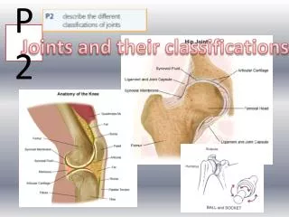
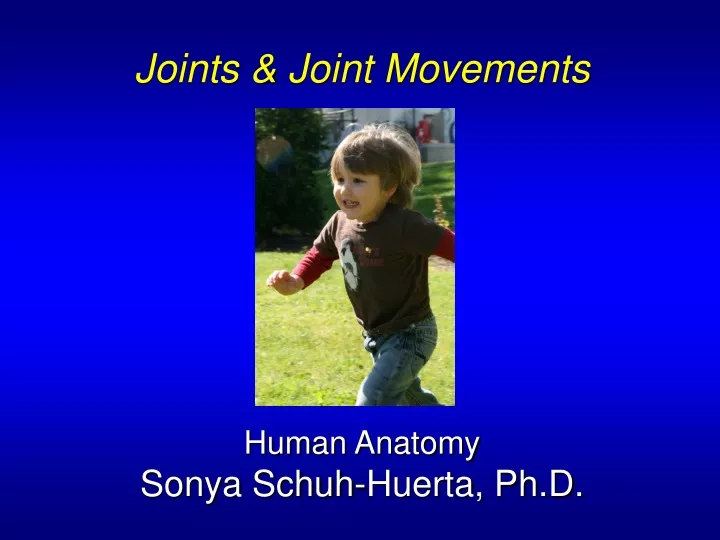
Joints & Joint Movements Human Anatomy Sonya Schuh-Huerta, Ph.D. Joints. Rigid elements of the skeleton meet at joints or articulations Greek root “arthro” means joint Structure of joints Enables resistance to crushing, tearing, & other forces. Classifications of Joints.

E N D
Joints & Joint Movements Human Anatomy Sonya Schuh-Huerta, Ph.D.
Joints • Rigid elements of the skeleton meet at joints or articulations • Greek root “arthro” means joint • Structure of joints • Enables resistance to crushing, tearing, & other forces
Classifications of Joints • Joints can be classified by function or structure • Functional classification based on amount of movement • Synarthroses immovable; common in axial skeleton • Amphiarthroses slightly movable; common in axial skeleton • Diarthroses freely movable; common in appendicular skeleton (all synovial joints)
Classifications of Joints • Structural classification based on • Material that binds bones together • Presence or absence of a joint cavity • Structural classifications include: • Fibrous • Cartilaginous • Synovial
Sutures – A Type of Fibrous Joint • Bones are tightly bound by a minimal amount of fibrous tissue • Only occur between the bones of the skull • Allow bone growth so the skull can expand with brain during childhood • Fibrous tissue ossifies in middle age • Synostoses = closed sutures
Syndesmoses – A Type of Fibrous Joint • Bones are connected exclusively by ligaments • Amount of movement depends on length of fibers • Tibiofibular joint = immovable synarthrosis • Interosseous membrane between radius & ulna = freely movable diarthrosis
Gomphoses – A Type of Fibrous Joint • Tooth in a socket • Connecting ligament the periodontal ligament
Fibrous Joints (a) Suture (b) Syndesmosis (c) Gomphosis Joint held together with very short, interconnecting fibers, and bone edges interlock. Found only in the skull. Joint held together by a ligament. Fibrous tissue can vary in length but is longer than in sutures. Peg-in-socket fibrous joint. Periodontal ligament holds tooth in socket. Socket of alveolar process Fibula Suture line Tibia Root of tooth Dense fibrous connective tissue Ligament Periodontal ligament
Cartilaginous Joints • Bones are united by cartilage • Lack a joint cavity • 2 types • Synchondroses • Symphyses
Synchondroses • Hyaline cartilage unites bones • Epiphyseal plates • Joint between first rib & manubrium (a) Synchondroses Bones united by hyaline cartilage Sternum (manubrium) Epiphyseal plate (temporary hyaline cartilage joint) Joint between first rib and sternum (immovable)
Symphyses • Fibrocartilage unites bones; resists tension & compression • Slightly movable joints that provide strength with flexibility • Intervertebral discs • Pubic symphysis • Hyaline cartilage present as articular cartilage
Symphyses (b) Symphyses Bones united by fibrocartilage Body of vertebra Fibrocartilaginous intervertebral disc Hyaline cartilage Pubic symphysis
Synovial Joints • Most movable type of joint!!! • All are diarthroses what does that mean? • Each contains a fluid-filled joint cavity
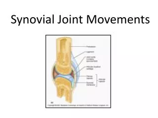
General Structure of Synovial Joints • Articular cartilage • Ends of opposing bones are covered with hyaline cartilage • Absorbs compression • Joint cavity (= synovial cavity) • Unique to synovial joints • Cavity is a potential space that holds a small amount of synovial fluid
General Structure of Synovial Joints • Articular capsule joint cavity is enclosed in a 2-layered capsule • Fibrous capsule dense irregular connective tissue, which strengthens joint • Synovial membrane loose connective tissue • Lines joint capsule & covers internal joint surfaces • Makes synovial fluid
General Structure of Synovial Joints • Synovial fluid • A viscous fluid similar to raw egg white • A filtrate of blood • Arises from capillaries in synovial membrane • Contains glycoprotein molecules secreted by fibroblasts
General Structure of Synovial Joints • Reinforcing ligaments • Often are thickened parts of the fibrous capsule • Sometimes are extracapsular ligaments located outside the capsule • Sometimes are intracapsular ligaments located internal to the capsule
General Structure of Synovial Joints Ligament Joint cavity (contains synovial fluid) Articular (hyaline) cartilage Fibrous capsule Articular capsule Synovial membrane Periosteum (a) A typical synovial joint
General Structure of Synovial Joints • Richly supplied with sensory nerves • Detect pain • Most monitor how much the capsule is being stretched
General Structure of Synovial Joints • Have a rich blood supply • Most supply the synovial membrane • Extensive capillary beds produce basis of synovial fluid • Branches of several major nerves & blood vessels
Synovial Joints with Articular Discs • Some synovial joints contain an articular disc • Temporomandibular joint & Knee joint • Occur in joints whose articulating bones have somewhat different shapes
How Synovial Joints Function • Synovial joints lubricating devices • Friction could overheat & destroy joint tissue & bone ends • Are subjected to compressive forces • Fluid is squeezed out as opposing cartilages touch • Cartilages ride on the slippery film
Bursae & Tendon Sheaths • Bursae & tendon sheaths are not synovial joints • Closed bags of lubricant • Reduce friction between body elements • Bursa = a flattened fibrous sac lined by a synovial membrane • Tendon sheath = an elongated bursa that wraps around a tendon
Bursae & Tendon Sheaths Coracoacromial ligament Acromion of scapula Subacromial bursa Coracoacromial ligament Joint cavity containing synovial fluid Subacromial bursa Cavity in bursa containing synovial fluid Fibrous articular capsule Humerus resting Hyaline cartilage Bursa rolls and lessens friction. Tendon sheath Synovial membrane Tendon of long head of biceps brachii muscle Fibrous capsule Humerus head rolls medially as arm abducts. Humerus Humerus moving (a) Frontal section through the right shoulder joint (b) Enlargement of (a), showing how a bursa eliminates friction where a ligament (or other structure) would rub against a bone
Movements Allowed by Synovial Joints • 3 basic types of movement • Gliding one bone across the surface of another • Angular movement movements change the angle between bones • Rotation movement around a bone's long axis
Gliding (a) Gliding movements at the wrist Gliding Joints • Flat surfaces of two bones slip across each other • Gliding occurs between: • Carpals • Articular processes of vertebrae • Tarsals
Angular Movements • Increase or decrease angle between bones • Movements involve: • Flexion & extension • Abduction & adduction • Circumduction
Angular Movements Extension Flexion (b) Angular movements: flexion & extension of the neck
Angular Movements Extension Flexion (c) Angular movements: flexion & extension of the trunk
Angular Movements Flexion Extension Flexion Extension (d) Angular movements: flexion & extension at the shoulder and knee
Angular Movements Abduction Circumduction Adduction (e) Angular movements: abduction, adduction, & circumduction of the upper limb at the shoulder
Rotation • Involves turning movement of a bone around its long axis • The only movement allowed between atlas & axis vertebrae • Occurs at the neck, shoulder, elbow, hip
Rotation Rotation Lateral rotation Medial rotation (f) Rotation of the head, neck, & lower limb
Special Movements • Elevation lifting a body part superiorly • Depression moving the elevated part inferiorly Elevation of mandible Depression of mandible Elevation Depression
Special Movements • Protraction nonangular movement anteriorly • Retraction nonangular movement posteriorly Protraction of mandible Retraction of mandible Protraction Moving a body part in the anterior direction Retraction Moving a body part in the posterior direction
Special Movements • Supination forearm rotates laterally, palm faces anteriorly • Pronation forearm rotates medially, palm faces posteriorly • Brings radius across the ulna
Special Movements Pronation (radius rotates over ulna) Supination (radius and ulna are parallel) Pronation (P) Rotating the forearm so the palm faces posteriorly Supination (S) Rotating the forearm so the palm faces anteriorly
Special Movements • Opposition thumb moves across the palm to touch the tips of other fingers Opposition
Special Movements • Inversion & eversion • Special movements at the foot • Inversion turns sole medially • Eversion turns sole laterally
Special Movements Inversion Eversion Inversion Turning the sole of the foot medially Eversion Turning the sole of the foot laterally
Special Movements • Dorsiflexion&plantarflexion • Up-and-down movements of the foot • Dorsiflexion lifting the foot so its superior surface approaches the shin • Plantar flexion depressing the foot, elevating the heel (ballet toe point)
Special Movements Dorsiflexion Plantar flexion Dorsiflexion Lifting the foot so its superior surface approaches the shin Plantar flexion Depressing the foot elevating the heel
Eversion of Ankle joint (5a) PLAY
Synovial Joints Classified by Shape • Plane joint • Articular surfaces are flat planes • Short gliding movements are allowed • Intertarsal & intercarpal joints • Movements are nonaxial • Gliding does not involve rotation around any axis
Plane Joint Nonaxial movement Metacarpals Carpals Gliding (a) Plane joint
Synovial Joints Classified by Shape • Hinge joints • Cylindrical end of one bone fits into a trough on another bone • Angular movement is allowed in one plane • Elbow, ankle, & joints between phalanges • Movement is uniaxial allows movement around one axis only
Hinge Joint Uniaxial movement Humerus Medial/ lateral axis Ulna Flexion & extension (b) Hinge joint
Synovial Joints Classified by Shape • Pivot joints • Classified as uniaxial – rotating bone only turns around its long axis • Examples • Proximal radioulnar joint • Joint between atlas & axis