Download
1 / 30
300 likes | 318 Views
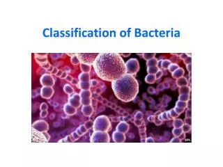
Classification of bacteria DR.THAMINA SAYYED REGISTRAR MICROBIOLOGY KKUH. Bacterial cells. Classification System. 3 Domains 1978 Carl Woese 1. Bacteria

E N D
Classification of bacteria DR.THAMINA SAYYED REGISTRAR MICROBIOLOGY KKUH
Classification System • 3 Domains 1978 Carl Woese • 1. Bacteria • Unicellular prokaryotes with cell wall containing peptidoglycan • 2. Archaea • Unicellular prokaryotes with no peptodoglycan in cell wall • 3.Eukarya • Protista • Fungi • Plantae • Animalia
Taxonomic Classification Categories • arranged in hierarchical order • species is basic unit Domain Kingdom Phylum or Division Class Order Family Genus Species
Prokaryote Classification • Technologies used to characterize and ID prokaryotes • microscopic examination • culture characteristics • biochemical testing • nucleic acid analysis • combination of the above is most accurate
Phenotypic Characteristics for Identifying Prokaryotes • often does not require sophisticated equipment • can easily be done anywhere
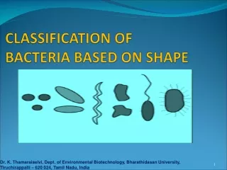
Microscopic Phenotypic Exam • size and shape and arrangement • enough information for diagnosis of certain infections • Gram stain • distinguishes between Gram + and Gram – bacteria • narrows the possibilities quickly
Microscopic Phenotypic Exam • special stain • allows for the distinction of microorganisms with unique characteristics • capsule • acid fast staining detects the waxy presence of Mycobacterium tuberculosis Capsule staining Acid fast staining of M. tuberculosis
CELL WALL Gram positive cell wall Gram negative cell wall • Consists of • a thick, homogenous sheath of peptidoglycan 20-80 nm thick • tightly bound acidic polysaccharides, including teichoic acid and lipoteichoic acid • cell membrane • Retain crystal violet and stain purple • Consists of • an outer membrane containing lipopolysaccharide (LPS) • thin shell of peptidoglycan • periplasmic space • inner membrane • Lose crystal violet and stain pink from safranin counterstain 11
Gram Positive Gram Negative 12
The Gram Stain Gram's Crystal iodine violet Decolorise with acetone Gram-positives appear purple Counterstain with e.g. methyl red Gram-negatives 13 appear pink
Gram-positive rods Gram-positive cocci Gram-negative rods Gram-negative cocci 15
Metabolic Phenotypic Exam • cultural approaches • required for positive diagnosis of infection • isolation and ID of pathogen • accuracy, reliability, and speed • methods used include • culture characteristics • biochemical reactions process
Serological Testing Phenotypic Exam • serological testing uses ELISA testing • fast and easy to use
Classification of medically significant bacteria • I.Thick rigid walled cells A. Free living extracellular 1.Gram positive a.CocciStaphylococcus - abcess Streptococcus - puemonia, Pharyngitiscellulitis b.Spore forming rods Aerobic Bacillus - Anthrax Anaerobic Clostridium - tetanus,gas gangrene botulism
c.Non spore forming rods (GRAM POSTIVE CONTD) 1-Non filamentous Cornybacterium – Diphtheria Listeria - meningitis 2.Filamentous Actinomycetes – Actinomycosis Nocardia - Nocardiosis
2.Gram negative A.Cocci Neisseria -Gonorrhoea, meningitis B.Rods 1.Facultative a. Straight 1.Respiratory org. Haemophillus- meningitis Bordatella-Whooping cough Legionella- Pneumonia 2.Zoonotic Brucella – Brucallosis Francisella –Tularemia Pasteurella –Cellulitis Yersinia - Plague
3.enteric & related (GRAM NEGATIVE CONTD) E.coli - UTI,Diarrhoea Enterobacter – UTI Serratia – Pneumonia Klebsiella – Pneumonia.UTI Salmonella – enterocolitis,typhoid fever Shigella – Enterocolitis Proteus – UTI b. Curved Campylobacter – Entericolitis helicobacter – Gastritis,Peptic ulcer Vibrio - Cholera
(Gram negative) • C.Aerobic Pseudomonas – pneumonia,UTI • D. Anaerobic Bacteroids – peritonitis 3.ACID FAST MYCOBACTERIUM - Tuberculosis & Leprosy
B . Non free living obligate intracellular parasites 1.Rickettsia – Rocky mountain spotted fever Typhus, Q fever 2.Chlamydia urethritis, trachoma. Psittacosis
Flexible thin walled Spirochaetes - Treponema – Syphilis Borrelia – Lyme disease Leptospira - leptospirosis Wall- less cells Mycoplasma - pneumonia
Subtyping & Its applications • To distinguishinguish between strains of different species • Biotyping • Serotyping • Antimicrobial susceptibility system • Bacteriophage typing • Bacteriocin typing
Genotypic Characteristics for Identifying Prokaryotes • the use of genotypic testing has increased with the availability of technology • genotypic testing is particularly useful in the case of organisms that are difficult to identify • several techniques include • gene probes • PCR • sequencing rRNA
gene probes • single stranded DNA that has been labeled with a identifiable tag, such as a fluorescent dye • are complementary to target nucleotide sequences • unique in DNA of pathogen
Genotypic Characteristics used in Classifying Prokaryotes( non culture methods) • PCR: polymerase chain reaction • used to detect small amounts of DNA present in a sample (blood, food, soil) • the PCR chain reaction is used to amplify the amount of DNA present • sequencing ribosomal RNA • of particular use for identifying prokaryotes impossible to grow in a culture • focus is place on the 16S molecules of the RNA because of it’s size • approximately 1500 nucleotides • once the 16S molecule is sequenced, it can then be compared to the sequences of known organisms
Genotypic Characteristics used in Classifying Prokaryotes • comparison of nucleotide sequences • differences in DNA sequence can assist in determination of divergence of evolutionary path for organisms • DNA hybridization • single strands of DNA anneal • 16S ribonucleic acid • comparing sequence of ribosomal RNA • relatedness to other organisms can be determined using numerical taxonomy • determined by the percentage of characteristics two organisms have in common