Download
1 / 31
460 likes | 1.04k Views
Classification of Bacteria. Bacteria are…. Prokaryotic Eukaryotic Can be Both. Prokaryotic (no nucleus)! Bacteria are the ONLY prokaryotes on Earth!. Bacteria are…. Unicellular Multicellular Can be Both. Unicellular! Bacteria are composed of a single cell!.

E N D
Bacteria are… • Prokaryotic • Eukaryotic • Can be Both Prokaryotic (no nucleus)! Bacteria are the ONLY prokaryotes on Earth!
Bacteria are… • Unicellular • Multicellular • Can be Both Unicellular! Bacteria are composed of a single cell!
In what kingdoms can we find bacteria? • Protista • Fungi • All of the above • None of the above Bacteria can be found in the kingdoms Archaebacteria & Eubacteria!
Bacteria • Prokaryotic – no nucleus & no membrane-bound organelles • Unicellular • Classified into 2 domains/kingdoms based on their cell walls & habitat • Kingdom Archaebacteria • Kingdom Eubacteria (means true bacteria)
Remember! The 3 Domain System • Reflects a greater understanding of evolution & molecular evidence • Domain Bacteria: eubacteria • Domain Archaea: archaebacteria • Domain Eukarya: plants, animals, protists, & fungi
Archaebacteria • In their own domain, Archaea, because they are so molecularly different from true bacteria! • They’re extremophiles – live in extreme environments (no oxygen, high salt, high temperatures, etc.) • Their cell walls do not contain peptidoglycan. • Obtain energy from the sun or inorganic molecules
Eubacteria • In the domain Bacteria • Live all over the world, around, in & on organisms • Their cell walls are made of peptidoglycan, a structural carbohydrate.
Bacteria Structures • Plasma Membrane: selectively permeable for exchange of nutrients and waste • Cytoplasm: fluid inside of the cell • Cell Wall: made of peptidoglycan in eubacteria
Bacteria Structures • DNA: clustered in a “nucleoid region” • Pili: small protein extensions used to anchor themselves OR sex pili aid in exchanging DNA • Flagella: tail used for movement • Ribosomes: translate genetic material into proteins
Bacteria Structures • Capsule: some have a sticky, gelatinous layer around the cell wall These bacteria are more likely to cause disease, because the capsule helps them to attach to host tissues.
Genetic Information in Bacteria • Chromosomal DNA: single DNA molecule arranged as a circular chromosome • Not enclosed in a nucleus! • Plasmid: small, circular chromosome piece containing only a few genes • Separate from the chromosomal DNA!
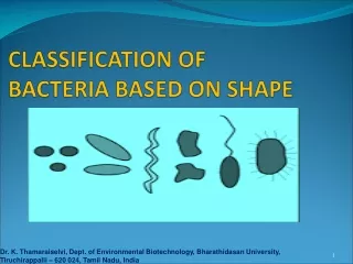
Scientists use several characteristics to classify bacteria. • Shape • Cell Wall • Metabolism
Classification by Shape • Coccus (cocci, pl.): round, can be found in clumps or lines • Baccillus (baccilli, pl.): rod shaped • Spirillum (spirilla, pl.): corkscrew (spiral) shaped
Spirillum Coccus Bacillus
Bacterial Arrangement • Singular • Pairs: Diplo- • Chains: Strepto- • Clusters: Staphylo- Diplococcus Staphylococcus Streptococcus
Streptococcus pyogenes, flesh-eating bacteria • Singular • Stapyhlo- • Strepto- • Diplo-
Staphylococcus aureus, common cause of skin infection, respiratory disease, & food poisoning. • Singular • Stapyhlo- • Strepto- • Diplo-
Neisseria meningitidis, diplococcus bacteria that causes meningitis • Singular • Stapyhlo- • Strepto- • Diplo-
Classification by Cell Wall • Eubacteria can be identified by the thickness of their cell wall with a process called gram staining. • The dye stains the peptidoglycan in the cell wall. • Can be used to identify types of eubacteria • Gram + Bacteria: stains purple; have a thick cell wall • Gram - Bacteria: stains pink; have a thin cell wall Gram Negative (-) Gram Positive (+)
outer membrane of lipopolysaccharides Gram-negative bacteria Gram-positive bacteria peptide side chains outer membrane cell wall peptidoglycan cell wall peptidoglycan plasma membrane plasma membrane protein Peptidoglycan: polysaccharides + amino acid chainsLipopolysaccharides: lipids + polysaccharides
Gram + • Gram + • Gram – • Both • Neither Bacillus anthracis
Thin cell wall of peptidoglyan • Thin cell wall • Thick cell wall • Both • Neither Spirillumvolutans
Gram + and Gram - • Gram + • Gram – • Both • Neither Staphylococcus aureus& Escherichia coli
A scientist uses gram staining on a colony of bacteria that she collected from a hot spring. What will her results look like? Neither! The bacteria collected was Archaebacteria. Gram staining will be ineffective, because Archaebacteria do not have peptidoglycan in their cell walls. Gram staining can only be used to identify Eubacteria! • Gram + • Gram – • Both • Neither
How would we classify these bacteria? Streptobacillus, Gram + Bacillus, Gram - Staphylococcus, Gram +
Classification By Metabolism • Bacteria can be classified by how they obtain energy & use it. • Heterotrophs: obtain food from another source • Autotrophs: make their own food • Chemoautotrophs: make their own food with inorganic molecules • Photoautotrophs: make their food using light
Metabolism & Respiration – Using their Food • Obligate aerobes: require O2 supply • Tuberculosis mycobacterium • Obligate anaerobes: cannot grow in the presence of O2 • Clostridium botulinum • Facultative anaerobes: do not require O2, but are not harmed by it (can live anywhere) • Escherichia coli
What do you think the dirtiest part of the classroom is? • Your group will swab a location of your choice and see if bacteria are present. • We will check the growth in a few days.
Directions to Prepare the Petri Dish • Pre-Lab Questions! • Write your group’s info. on the bottom of the petri dish. • Initials of all members • Class period • Agree on a location to test & come to me for a sterile Q-tip. • Rub 1 side of the Q-tip on the location, then lightly rub the same side onto the agar. • Finish your drawings on your lab handout. • Replace the lid & tape it shut. • Place it on the tray.