Download
1 / 45
450 likes | 606 Views
Today’s Plan Sept 10, 2013. Bellwork bonemarkings On your own paper: id functions of the skeletal system. Classwork Human Skeleton: General skeleton, classification of bones Axial vs. Appendicular Words to describe bone structures Skull Assign skull project 15 minute skull prep time.

E N D
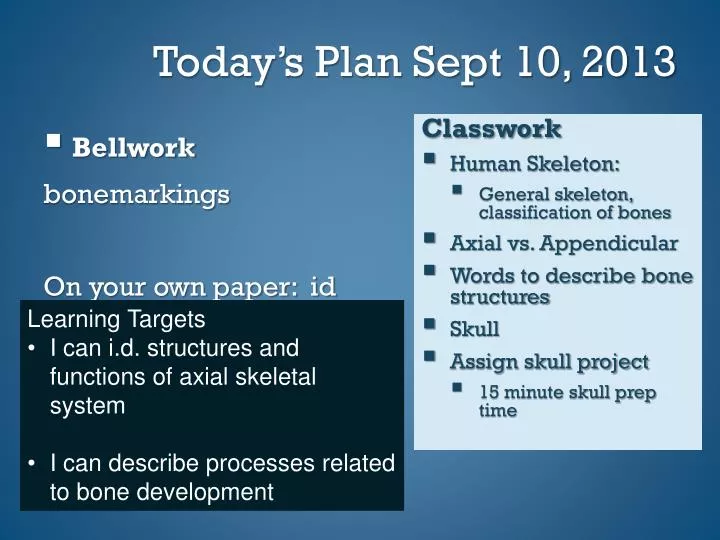
Today’s Plan Sept 10, 2013 • Bellwork bonemarkings On your own paper: id functions of the skeletal system. Classwork • Human Skeleton: • General skeleton, classification of bones • Axial vs. Appendicular • Words to describe bone structures • Skull • Assign skull project • 15 minute skull prep time • Learning Targets • I can i.d. structures and functions of axial skeletal system • I can describe processes related to bone development
Today’s Plan Sept 11, 2013 • Bellwork bonemarkings On your own paper: id functions of the skeletal system. Classwork • Human Skeleton: • Bone Development • Skull labeling • 15 minute skull prep time • Learning Targets • I can i.d. structures and functions of axial skeletal system • I can describe processes related to bone development
Today’s Plan Sept 12, 2013 • Bellwork Concept map and label skull bones On your own paper: id functions of the skeletal system. Classwork • Human Skeleton: • Skull practice • Skull models 20 minute skull prep time • Learning Targets • I can i.d. structures and functions of axial skeletal system • I can describe processes related to bone development
Functions of Skeletal System • Support Lower limbs, pelvis, vertebrae support upper body. • Body Movement/ Leverage Bones and muscles act as levers • Protection Eyes, ears bones, brain, heart, lungs
Functions of Skeletal System 4. Blood Cell production • Hemopoiesis • Begins in yolk sac then to liver and spleen and eventually bone marrow • 2 kinds • Red marrow • Platelets, RBC and WBC • Infants mostly have red marrow • Adults: red marrow is stored in skull, ribs, sternum, clavicles, vertebrae and pelvis • Yellow Marrow • Stores fat • Within long bones
Functions 5. Inorganic salt storage • Calcium • Blood clot formation, muscle contraction • Nerve impulses • Phosphorus • Mg • Na • K
Two divisions of skeleton • the axial and the appendicular skeleton. • Axial: includes all bones that support organs of the head, neck, and trunk. • 40% of skeleton Axial Skeleton
Skull :cranium and facial bones • Middle Ear Bones (3 per ear) • Hyoid bone • Vertebral column • Thoracic cage: ribs and sternum Axial Skeleton Major Components
The appendicular skeleton consists of the bones of the limbs and bones that anchor the limbs to the axial skeleton. • Pectoral girdle: scapula, clavicle. • Upper limbs: humerus, radium, ulna, carpals, metacarpals, phalanges. • Pelvic girdle:coxal bones. • Lower limbs: femur, tibia, fibula, patella, tarsals, metatarsals, phalanges. Appendicular Skeleton
Classification of Bones *Bones are classified according to shape* 1. Long Bones • Consists of a shaft with 2 ends • Humerus, femur, radius, ulna, palms, soles • Short Bones • Cube-like shaped bones • Carpals, tarsals
3. Flat Bones • Thin and usually curved bones • Skull bones, sternum, scapula, ribs • Irregular Bones • Bones that not are long, short or flat • Vertebrae, auditory ossicles
Sesamoid Bones • Usually small, round, and flat. They develop inside tendons • Patella • Wormian (sutural) Bones • Tiny bones in skull that lie between major skull bones
Depressions and openings allowing blood vessels and nerves to pass Meatus – Canal-like passageway • auditory
Sinus – cavity within bone, filled with air and lined with mucous membrane • skull
Fossa – shallow, basin like depression in a bone, often serving as an articular surface • Pelvis, scapula, mandibular
Groove – Furrow • Mandibular groove
Suture • Interlocking union b/t bones
Condyles • Rounded projections • Usually articular surfaces
Frontal bone: forehead • Parietal bones: top of the skull • Occipital bone: back of the skull • Temporal bones: side of skull, near ears • Sphenoid bone:base of the cranium • Ethmoid bone: roof of the nasal cavity Cranium
Maxillary bones: upper jaw, hard palate • Palatine bones: hard palate, nasal cavity • Zygomatic bones: cheek bones • Lacrimal bones: orbit of the eye • Nasal bones: bridge of the nose • Vomer bone: nasal septum • Nasal conchae: walls of the nasal cavity • Mandible: lower jaw Facial Skeleton
The skull at birth is not fully developed. • Fibrous membranes, fontanels, connect the cranial bones. • The fontanels allow movement of the bones to enable the skull to pass through the birth canal. • The fontanels close as cranial bones grow. Infantile Skull
Frontal • Spenoidal • Ethmoidal • maxillary Sinuses
Cervical vertebrae: seven vertebrae of the neck, includes atlas and axis • Thoracic vertebrae: twelve vertebrae that articulate with the ribs • Lumbar vertebrae: five vertebrae that make up the small of the back Vertebral Column
Sacrum: five vertebrae that fuse in early adulthood, part of the pelvis • Coccyx: four small fused vertebrae Vertebral Column Figure 7.34
Ribs: twelve pair of ribs attached to each thoracic vertebrae. • Seven pairs: true ribs and attach to the sternum by costal cartilage. • Two pairs: false ribs that attach to cartilage. Thoracic Cage
Two pairs: floating ribs that do not attach to the sternum or its cartilage. • Sternum: the manubrium, the body, and the xyphoid process. Thoracic Cage
http://highered.mcgraw-hill.com/sites/0072919329/student_view0/chapter7/labeling_exercises.html#http://highered.mcgraw-hill.com/sites/0072919329/student_view0/chapter7/labeling_exercises.html#