Download
1 / 72
840 likes | 2.18k Views
FILARIASIS. Species. Geographic distribution. Pathogenicity. Adults (site of infection). Microfilariae (characteristics. Vector. Wuchereria bancrofti. Asia, Pacific, Tropical Africa, Americas. Lymphagitis, fever, elephantiasis hydrocoele, chyluria. Lymphatics.

E N D
FILARIASIS para-lab by l. wafa menawi
Species Geographic distribution Pathogenicity Adults (site of infection) Microfilariae (characteristics Vector Wuchereria bancrofti Asia, Pacific, Tropical Africa, Americas Lymphagitis, fever, elephantiasis hydrocoele, chyluria Lymphatics Found in blood, sheathed, periodicity variable Culicidae (mosquitoes) Brugia malayi South and East Asia Lymphagitis, fever. Elephantiasis Lymphatics Found in blood, sheathed, nocturnally periodic or subperiodic Culicidae (mosquitoes) Dipetalonema perstans Africa and South America No definite pathogenicity Peritoneal & pleural cavity Found in blood, unsheathed, nocturnally subperiodic Culicoides (biting midges) Dipetalonema streptocerca Africa (Ghana and Congo) Cutaneous oedema, elephantiasis Subcutaneous tissues Found in skin, unsheathed, nonperiodic Culicoides (biting midges) Mansonella ozzardi Central and South America No definite pathogenitis Peritoneal cavity Found in blood, unsheathed, nonperiodic Culicoides (biting midges) Loa loa Tropical Africa Skin swellings, allergic reactions Subcutaneous tissues Found in blood, sheathed, diurnally periodic Chrysops (Tabanidae or Horse fly) Onchocerca volvulus Africa, Central and South America Skin nodules, occular complications (blindness) Subcutaneous tissues Found in skin, unsheathed, nonperiodic Simulium (Black fly)
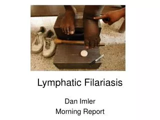
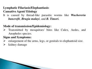
Lymphatic filariasis • Wuchereriabancrofti and Brugiamalayi are filarial nematodes • Spread by several species of night - feeding mosquitoes • Causes lymphatic filariasis, also known as Elephantiasis • Commonly and incorrectly referred to as “Elephantitis” para-lab by l. wafa menawi
Definitive host • Humans are the definitive host for the worms that cause lymphatic filariasis • There are no known reservoirs for W.bancrofti. • B.malayi has been found in macaques, leaf monkeys, cats and civet cats para-lab by l. wafa menawi
Anopheles Intermediate host • W.bancroftiis transmitted by Culex, Aedes, and Anopheles species • B.malayi is transmitted by Anopheles and Mansonia species. Aedes Culex Mansonia para-lab by l. wafa menawi
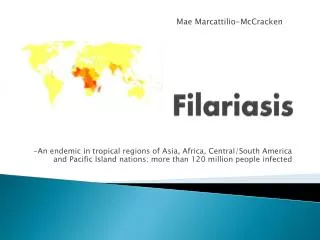
Epidemeology • Endemic in 83 countries • 1.2 billion at risk • More than 120 million people infected • More than 25 million men suffer from genital symptoms • More than 15 million people suffer from lymphoedema or elephantiasis of the leg para-lab by l. wafa menawi
Morphology • Adult: White and thread-like. Two rings of small papillae on the head. • Female:5~10cm in length • Male: 2.5~4cm and a curved tail with two copulatory spicules. para-lab by l. wafa menawi
Microfilaria: 177~296 µm in length, a sheath with free endings. Bluntly rounded anteriorly and tapers to a point posteriorly. A nerve ring with no nuclei at anterior 1/5 of the body. para-lab by l. wafa menawi Wuchereria bancrofti Brugia malayi
B.malayi microfilariae are slightly smaller than those of W.bancrofti. • Microfilariae are sheathed, and about 200 to 275 µm. • Not much is known about the adult worms, as they are not often recovered • One distinctive feature of B.malayi is that the microfilarial nuclei extends to the tip of the tail para-lab by l. wafa menawi
Anterior part and posterior part of bancroftian microfilaria
Bancroftianmicrofilaria:body nuclei equal sized, clearly, defined, countable without caudal nucleus
Microfilaria of B. malayi: the body nuclei is unequal sized, coalescing,uncountable. The cephalic space is longer with two caudal nuclei.
Species Size of Microfilariae Morphology of microfilariae Wuchereria bancrofti 210 – 320m by 8 - 10m Sheathed. Tail pointed and clear Brugia malayi 170 – 260m by 5 - 6m Sheathed. Tail pointed with 2 nuclei Loa loa 230 – 300m by 6 - 8m Sheathed. Tail blunt with nuclei Mansonella perstans 200m by 6m Unsheathed. Tail blunt with nuclei Mansonella ozzardi 250m by 6 - 7m Unsheathed. Tail pointed and clear
Life cycle • Host: Mosqutoes (intermediate host) Human (final host) • Location: Lymphatics and lymph nodes • Infective stage: Infective larvae • Transmission stage: Microfilariae • Diagnostic stage: Microfilariae para-lab by l. wafa menawi
Life cycle para-lab by l. wafa menawi
Wuchereria Life Cycle para-lab by l. wafa menawi
A large number of 3rd stage larvae of a filarial sp emerging from the proboscis of a mosquito.
Nocturnal periodicity • Phenomen which the number of microfilariae in peripherial blood is very low density during daytime, but increase from evening to midnight and reach the greatest density at 10p.m to 2 a.m. • May be related to cerebral activity and vasoactivity of pulmonary vessels. para-lab by l. wafa menawi
Larva deposited by mosquito bite • Travel through dermis to lymphatic vessels • Growth (approx 9 months) to mature worms(20-100mm long) • Worms live 5-7 years (occasionally up to15 years) • Mate->Microfilariae (1st stage larva) • Females->release up to 10,000 microfilariae/day into bloodstream • Microfilarie taken up by mosquito bite • Develop into 2nd and 3rd stage larva over 10-14 days inside mosquito vector para-lab by l. wafa menawi
Clinical features • Initially asymptomatic • Symptoms develop with increasing numbers of worms • Less than 1/3 of infected individuals have acute symptoms • Clinical Course is 3 phases: • Asymptomatic Microfilaremia • Acute Adenolymphangitis (ADL) • Chronic/Irreversible lymphedema • Superimposed upon repeated episodes of ADL para-lab by l. wafa menawi
Presents with sudden onset of fever and painful lymphadenopathy • Retrograde Lymphangitis • Inflammation spreads distally away from lymph node group • Immune mediated response to dying worms • Most common areas: Inguinal nodes and Lower extremities para-lab by l. wafa menawi
Inflammation spontaneously resolve after 4-7 days but can recur frequently • Recurrences usually 1-4 times/year with increasing severity of lymphedema • Secondary bacterial infections in edematous(elephantatic) areas • Filarial fever (fever w/o lymphangitis) • Tropical Pulmonary Eosinophilia • Hyperresponsiveness to microfilariae trapped in lungs • Nocturnal Wheezing para-lab by l. wafa menawi
Lymphedema Mostly LE and inguinal, but can affect UE and breast Initially pitting edema, with gradual hardening of tissues hyperpigmentation & hyperkeratosis GenitaliaHydroceles Chronic filariasis para-lab by l. wafa menawi
Renal involvement • Chylurialymph discharge into urine • Loss of fat and protein hypoproteinemia & anemia • Hematuria, proteinuria from ?immune complex nephritis • Secondary bacterial/fungal infections para-lab by l. wafa menawi
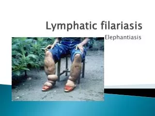
Elephantiasis: accumulation of lymph in extremeties, fibrosis, and thickening of skin. para-lab by l. wafa menawi
Elephantiasis due to Brugia malayi. Pitting does not occur in this stage (solid edema)
Elephantiasis due to Brugia malayi, complicated by severe dermatitis and secondary bacterial infection
This lady has elephantiasis of the right leg and edema in the left
The standard method for diagnosing active infection is the identification of microfilariae by microscopic examination However, microfilariae circulate nocturnally, making blood collection an issue Lab diagnosis para-lab by l. wafa menawi
A “card test” for parasite antigens requring only a small amount of blood has been developed • Does not require laboratory equipment • Blood drawn by finger stick • Urinalysis, CBC and Comprehensive Chemistries • Foot Biopsy: Normal Skin with areas of chronic inflammation para-lab by l. wafa menawi
Species Geographic location Periodicity Collection time Wuchereria bancrofti Tropics / Subtropics Nocturnal 12.midnight. Wuchereria bancrofti Pacific Diurnal subperiodic 16.00 hours Brugia malayi SE Asia and SW India Nocturnal 12.midnight. Brugia malayi Indonesia Nocturnal subperiodic 21.00 hours Brugia timori Indonesia Nocturnal 12.midnight Loa loa West / Central Africa Diurnal 13.00 hours Mansonella perstans Africa / S. America Non periodic Any time Mansonella ozzardi Central & S America Non periodic Any time
Microfilariae are seen in blood smears and are DIAGNOSTIC para-lab by l. wafa menawi
Blood Smear - Microfilaria • Note wavy microfilarial worm in the thick part of blood film. • Dark blue structures are nuclei • Tail end tapering (no nuclei) • Sheath covering worm. para-lab by l. wafa menawi