Download
1 / 37
370 likes | 439 Views
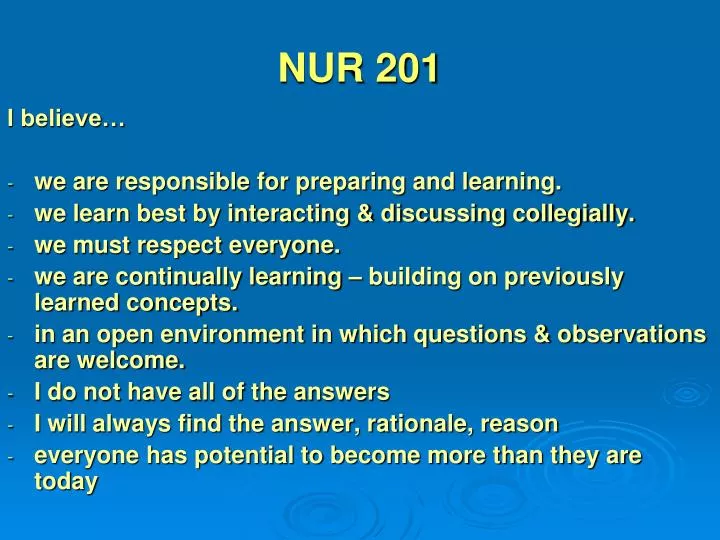
NUR 201. I believe… we are responsible for preparing and learning. we learn best by interacting & discussing collegially. we must respect everyone. we are continually learning – building on previously learned concepts. in an open environment in which questions & observations are welcome.

E N D
NUR 201 I believe… • we are responsible for preparing and learning. • we learn best by interacting & discussing collegially. • we must respect everyone. • we are continually learning – building on previously learned concepts. • in an open environment in which questions & observations are welcome. • I do not have all of the answers • I will always find the answer, rationale, reason • everyone has potential to become more than they are today
Interferences with VentilationObjectives • Discuss assessment—breath sounds • Describe diagnostic tests for pulmonary function • Discuss acid-base balance • Examine signs, symptoms, pathophysiology, treatments, and nursing care of respiratory distress syndromes • Discuss nursing interventions – mechanical ventilation, tracheostomy, postural drainage • Discuss pulmonary accidents—chest trauma, aspiration
Content Approach • Anatomy & Physiology Review • Demographics/occurrence • Pathophysiology • Clinical Picture • Medical Management • Nursing Process (APIE) Assessment - Nursing Actions - Education
Interferences with VentilationRespiratory Anatomy & Physiology • Anatomy • Structure of the Chest Wall: Ribs, pleura, muscles of respiration • Upper Respiratory: nose, pharynx, adenoids, tonsils, epiglottis, larynx, and trachea • Lower Respiratory: bronchi, bronchioles, alveolar ducts, and alveoli • Physiology • Ventilation: inspiration and expiration • Elastic Recoil: elastin fibers that recoil after expansion • Diffusion: Exchange of oxygen and carbon dioxide • Arterial Blood Gases / Oximetry
Interferences with VentilationAssessment • History • Cues to Respiratory Problems: • Shortness of breath – dyspnea • Orthopnea / Nocturnal dyspnea • Wheezing • Cough / sputum production • Hemoptysis • Voice change • Fatigue
Interferences with VentilationAssessment Thorax & Lungs • Inspection: • Posture, chest movement, abnormalities of sternum • Respiratory rate, depth, rhythm • Palpation: • Equality of chest expansion • Tactile Fremitus • Percussion: • Hyperresonance • Dullness • Auscultation: • Discontinuous: fine crackles/rales / coarse crackles / rales • Continuous: Wheeze, Rhonchi • Pleural friction rub
Respiratory AssessmentAssessment Definition Clinical Picture
Interferences with VentilationDiagnostic Studies • Blood Studies: Hgb, Hct, ABGs • Sputum Studies: C&S, Gram Stain, Acid-fast smear; Cytology • Radiology: • Chest x-ray-- posterior-anterior / lateral • Computed tomography (CT) – cross sections of the lung with and without contrast – used often • Magnetic resonance imaging (MRI) – images of pulmonary structures – limited use • Pulmonary angiogram – x-rays after injection of radiopaque dye– used to dx pulmonary embolism • Positron emission tomography (PET) – IV glucose administration – malignant tumors show increased uptake of glucose • Ventilation-Perfusion Scan – Perfusion: isotope administration which outlines pulmonary vasculature; Vent: inhalation of radioactive gas which outlines the alveoli – dx pulmonary emboli
Interferences with VentilationDiagnostic Studies • Endoscopic Exams (done in x-ray or OR): • Bronchoscopy – fiberoptic visualization of bronchi – biopsy; also used to remove mucous plugs, foreign bodies, obstructions • Mediastinoscopy – scope through a small incision n the suprasternal notch – visualize mediastinum for tumors, lymph nodes, infections, sarcoidosis • Biopsy: Transbronchial or open lung biopsy – done in x-ray or OR • Thoracentesis – insertion of a needle into the pleural space – pleural fluid, install medication - done at bedside • Pulmonary Function Testing – tests to measure lung volumes and used to dx pulmonary disease, monitor progress, evaluate disability, evaluate response to bronchodilators – done in pulmonary lab • Skin Testing – intradermal planning of test dose to assess skin reaction by measuring mm induration – TB, various lung diseases
Pulmonary Function Test Relationship of Lung Volumes & Capacities
Pair Share – Critical Thinking • Upon performing a lung sound assessment of the anterior chest, the nurse hears moderately loud sounds on inspiration that are equal in length with expiration. Where in the airway would this lung sound be considered normal? a. Trachea b. Primary bronchi c. Lung fields d. Larynx
Pair Share – Critical Thinking • The name that describes the particular lung sound in the previous questions is which of the following? • a. Bronchial • b. Bronchovesicular • c. Vesicular • d. Basilar
Interferences with VentilationRegulation of Acid-Base BalanceReview • Acid – contributes hydrogen ion • Two types: • Volatile respiratory acid • Dehydrates and excreted in the form of a gas • Nonvolatile metabolic acid • Metabolized and excreted in the form of body fluids
Interferences with VentilationRegulation of Acid-Base Balance Review • Base – accepts or removes hydrogen ion • Buffer- controls the hydrogen ion concentration: • Absorbing hydrogen ions when an acid is added OR • Releasing hydrogen ions when base is added. • Three Buffer Systems: • Bicarbonate – operates in lungs & kidneys • Phosphate – renal tubules • Protein – Hgb, plasma proteins, & intracellular protein
Interferences with VentilationRegulation of Acid-Base Balance • Factors to remember: • Lungs – Eliminate or retain carbon dioxide C02 • Kidneys – excrete or form bicarbonate HC03 Food – converted by the body – H20 + CO2 + energy Lung Kidney C02 + H20 = H2CO3 = HCO3- + H+
Interferences with VentilationRegulation of Acid-Base Balance • Lungs/Respiratory System • Increase or decrease hydrogen ion concentration • Through respiratory rate and depth • Result: C02 is either retained or eliminated • Changes can occur within minutes • Controlled in the medulla oblongata—respiratory center metabolic: > = increased; < = decreased • <pH causes > respirations = <C02 + correcting pH • >pH causes < respirations = >C02 + correcting pH
Interferences with VentilationRegulation of Acid-Base Balance • Renal System • Reabsorb and conserve bicarbonate • Can generate additional bicarbonate and eliminate excess hydrogen ions as compensation for acidosis • Three mechanisms: • Secretion of small amounts of free hydrogen into the renal tubule • Combination of hydrogen ions with ammonium to form ammonium • Excretion of weak acids • Urine pH 4 – 8
Interferences with VentilationRegulation of Acid-Base Balance> = increased; < = decreased
Acid-Base ImbalanceRespiratory Acidosis • Hypoventilation from primary lung problem • Atelectasis • Pneumonia • Respiratory failure • Airway obstruction • Chest wall injury • Cystic fibrosis • Hypoventilation from other factors • Drug overdose • Head injury • Paralysis of respiratory muscles • Obesity
Acid-Base ImbalanceRespiratory Alkalosis • Hyperventilation from primary lung problem • Asthma • Pneumonia • Inappropriate ventilator settings • Hyperventilation from other factors • Anxiety • Disorders of the central nervous system • Salicylate overdose
Interferences with VentilationRegulation of Acid-Base BalanceRespiratory Function
Pair Share – Critical Thinking • What acid-base imbalance would you suspect for the patient having respiratory problems with respiratory rate: 28/min and expiratory wheezing?
Interferences with VentilationRegulation of Acid-Base BalanceMetabolic Function