Download
1 / 25
250 likes | 257 Views
Evolving Technique: PFA in Young Patients – a Case Approach. Phil Davidson, MD Davidson Orthopaedics Park City, Utah Ortho Summit, Las Vegas Dec 6, 2018. Disclosures. none. Our Case: 38 year old Firefighter with anterior knee pain, now unable to manage ladders.

E N D
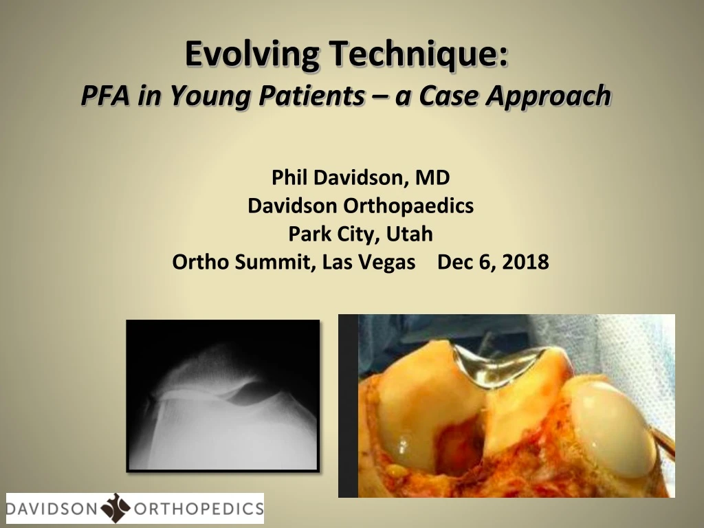
Evolving Technique:PFA in Young Patients – a Case Approach Phil Davidson, MD Davidson Orthopaedics Park City, Utah Ortho Summit, Las Vegas Dec 6, 2018
Disclosures none
Our Case: 38 year old Firefighter with anterior knee pain, now unable to manage ladders • She has already had PT, NSAID, Bracing and injections to include corticosteroid and HA • No trauma hx • Scope chondroplasty with no relief 2 yrs ago • Pain worse with descent • Pain exclusively in PF area
Radiography Merchant Xray- need dedicated board/jig >145 considered “shallow” Our patient: Sulcus angle: 132º Congruence angle: 30º
Radiography- Patellar Height • Caton-Deschamps (CD) Ratio (X/Y) • NL appx 0.6-1.3 • Very handy to use digital measuring tools • Patellar Alta and Baja
MRI Imaging – our patientTTTG 15mm (>18 TTO)CD ratio 1.1 (>1.3 Alta)
Audience Participation • Activity modification, bracing, more non surgical RX • TKA • Biological Resurfacing • TTO alone • PFA with prox realignment • PFA with TTO
How should we approach options here? • Etiology • Anatomy • Biologic vs Prosthetic
Etiology of PF degeneration • Traumatic (blow) • Malalignment • Morphology • Instability • Systemic DJD
The majority of isolated PF DJD in “younger patients” is associated with abnormal anatomy1. Abnormal Morphology2. Abnormal Geometry RotationHeightVersion3. Generalized Laxity 28 year old female
Morphology • Both patellar and trochlear morphology need to be identified in considering treatment options • Abnormal morphology can create stresses on repairs • Implant choice affected by trochlear and patellar shapes Wiberg Classification Dejour Classification
Geometry • Geometric alignment needs to be considered in 3-D • Patellar position M-L • Valgus knee • Patellar “tilt” • Femoral version • Patellar height • Correction targeted at specific malalignment/rotation
Limb Rotation – femur and tibia • Both Femoral Version AND Tibial Torsion bear on PF forces • Femoral Anteversion • NL female 13 • External Tibial Torsion • NL female 27
Extensor Realignment • Medial Plication • Lateral Lengthen, not release • Need “normal” tissue to plicate • i.e. not markedly lax • Easily incorporated into PFA “Selective” lateral release, preserving underlying synovial layer– part of realignment, not alone!
MPFL combo with PFA • This is indicated when DJD coexists with recurrent instability and/or laxity • Need to protect patellar implant • Avoid patellar bone tunnel techniques
TTO or Trochleoplasty with PFA • Medialization can correct for increased TT-TG or TT-PCL • Move proximal to address patellar baja • Distalize to address patellar alta
Biologic or Prosthetic Resurfacing ???? Key decision making points • Multifactoral decision • Lesion: focal or diffuse • Patient Factors • Comorbidities • Osteophytes, catabolic environment • Bipolar • Resources Available
Biological Options • Scaffolds • Cell Therapy • Osteochondral Grafts • Autogenous • Limited use • Allograft • Fresh stored • Cryopreserved • Cartilage Grafts • Minced, ground, lamellar • Cryopreserved • Non-viable (scaffold)
Onlay vs Inlay Joint Resurfacing - Patella • Inlay useful for focal defects and for “normal” morphology • Onlay needed for diffuse chondral disease or “abnormal” morphology • I use Onlay 98% of cases
Onlay vs Inlay Joint Resurfacing for FTG • Onlay device replaces anatomy, but may add unwanted volume • Inlay device based on ambient anatomy • Inlay device allows for concurrent realignment • Inlay device inherently stable • Inlay typically more anatomic • Inlay allows easier conversion to TKA Onlay Inlay
Operative Images and alignment. I did Prox Realignment with medial plication and lateral lengthening Extension Flexion