Download
1 / 35
360 likes | 816 Views
Porphyrias and errors in heme metabolism. Justin Mak Rubeena Patil Jonathan Mak Aisha Shoaib SEPTEMBER 28 TH 2012. PHM142 Fall 2012 Instructor: Dr. Jeffrey Henderson. Outline. Introduction Signs, Symptoms and Treatments Pathway of Heme Synthesis

E N D
Porphyrias and errors in heme metabolism Justin Mak RubeenaPatil Jonathan Mak Aisha Shoaib SEPTEMBER 28TH 2012 PHM142 Fall 2012 Instructor: Dr. Jeffrey Henderson
Outline • Introduction • Signs, Symptoms and Treatments • Pathway of Heme Synthesis • Emphasis on Porphyria cutanae tarda (PCT)
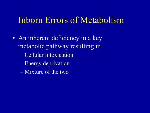
Introduction • Porphyrias are a set of diseases that result from enzyme deficiencies in the heme synthesis pathway. • Heme is mostly used for its incorporation into hemoglobin and role in red blood cells, but is also needed for cytochrome P450 function. • Each disease is associated with a deficiency in one of the eight enzymes in the pathway. • They fall under two categories based on their clinical symptoms: acute hepatic and erythropoietic porphyrias. Heme
Disease Mechanism • Generally, the disease mechanism for porphyrias is that their respective enzymatic deficiencies lead to the accumulation of porphyrins and porphyrin precursors that ultimately produce free radicals. • For acute hepatic porphyrias, delta-aminolaevulinic acid (ALA) and porphobilinogen (PBG) accumulate and produce free radicals via autoxidation. • For erythropoieticporphyrias, uroporphyrins, coproporphyrins, and protoporphyrins accumulate and produce free radicals via the absorption of visible light. • The generated free radicals participate in oxidative stress reactions, such as lipid oxidation and protein crosslinking, that lead to membrane and mitochondrial damage, ultimately promoting cell death. • Loss of negative feedback of heme leads to further accumulation of porphyrins.
Genetics • Enzyme deficiencies in porphyrias are inherited: acute intermittent porphyria (AIP), hereditary coproporphyria (HCP), variegate porphyria (VP), and erythropoietic protoporphyria (EPP) are autosomal dominant. • ALA dehydratase deficiency porphyria (ADP) and congenital erythropoietic porphyria (CEP) are autosomal recessive. • Porphyria cutanea tarda (PCT) can be autosomal dominant, but is mostly sporadic.
Environmental Factors • Due to incomplete penetrance, inheritance of an enzyme deficiency of an autosomal dominant porphyria does not necessarily lead to clinical symptoms. Symptoms of acute porphyrias tend to come in the form of "attacks" which may be induced by other genetic factors or environmental factors, such as agents that promote porphyrin and porphyrin precursor synthesis and/or agents that induce cytochrome P450s.
Symptoms of Cutaneous Forms • Occur most commonly with exposure to sunlight • Mainly skin symptoms that occur • Due to excess poryphorins that accumulate in surface of skin • Symptoms: • Fluid filled blisters • Changes in pigmentation • Breakdown (necrosis) of the skin when exposed to sunlight • Overall skin can become scarred, brown, blotchy and fragile
Symptoms of Acute Forms • Originate mainly in nervous system • Symptoms last around 1-2 weeks • Possible mechanisms include damage by free radicals, direct neurotoxicity of ALA acid, and the deficiency in nervous tissue Symptoms: • Severe abdominal pain • Muscle weakness and pain, tingling, or numbness and possibly paralysis • Pain in arms, legs, back • Constipation • Vomiting • Diarrhea • Insomnia • Seizures and Confusion • Anxiety and paranoia • Fever
Treatment for Cutaneous Forms • Avoiding sunlight • Attention to skin care • Beta-carotene supplements • Function to neutralize the effects of reactive protoporphyrins
Treatment for Acute Forms Several treatments can be used to control neurological symptoms and defective heme production: • Carbohydrate such as glucose • To help limit the synthesis of porphyrins • Phlebotomy (removal of blood) • To reduce excessive iron stores which improves heme synthesis • Sedatives to help with anxiety • Pain medications such as opiates • Hematin given intravenously • Hematin are heme-like substances that inhibit ALA synthase and the accumulation of toxic precursors
ALA Synthetase • Most important rate limiting enzyme • Deficiency may cause Sideroblasticanemia • Bone marrow produces ringed sideroblast? • X-linked • Respond to pyridoxine treatment
ALA dehydratase deficiency porphyria(DOSS porphyria) • Autosomal recessive • Very rare • Symptoms: Abdominal pain, neuropathy ALA dehydratase Aminolevulinic Acid Porphobilinogen
Acute intermittent porphyria (AIP) • 2nd most common form of porphyria • Caused by deficiency of PGB deaminase • Metabolite porphobilinogen accumulates in cytoplasm • Symptoms: • Localized abdominal pain • Urinary symptoms • Peripheral nerutopathy • raised concentration of urinary porphyrins • Treatment • Hematin, Hemearginate • Do not cure but reduces symptoms • Inhibit ALA synthase which occurs at the beginning of heme biosynthesis PGB deaminase Hydroxymethylbilane Porphobilinogen (PGB)
Congenital erythropoietic porphyria (CEP) • Deficiency of Uroporphyrinogen III synthase • Rare autosomal recessive (1 in 1,000,000) • Severe photosensitivity Uroporphyrinogen III synthase Hydroxymethylbilane Uroporphyrinogen III
Porphyria cutanae tarda (PCT) • Most common porphyria • Classified as such when Uroporphyrinogen decarboxylase activity <20% • Hepatic disorder • Inherited or obtained through Hepatitis C, drugs, alcohol, poisons • Treatment: discourage risk factors and treat symptoms; • can draw blood to reduce iron in the liver until the serum ferritin reaches normal iron levels. • Chloroquineor hydroxychloroquine can move excess porphyrins from the liver and promote excretion. Can be used when drawing blood is not recommended. • Avoid causes of PCT Uroporphyrinogen decarboxylase Coproporphyrinogen III Uroporphyrinogen III
Hereditary coproporphyria: • Deficiency of Coproporphyrinogen III Oxidase • Autosomal dominant • No cure exists
Variegate porphyria • Deficiency in protoporphyrinogen IX-oxidase • Autosomal dominant
Erthropoietic Protoporhyria • Caused by deficiency of Ferrochelatase • Autosomal dominant • Photosensitivity- can be managed by limiting exposure
Summary Slide A • Porphyria is caused by a deficiency in any of the 8 enzymes of heme synthesis. • Leading to the build up of heme precursors • Divided into two categories based on clinical symptoms: acute hepatic and erythropoeitic. • Enzymatic deficiencies are inherited, but symptoms may not appear unless induced by environmental agents. • Main symptoms associated: porphyrin accumulation in various locations, photosensitivity, pain, numbness, vomiting, and seizures. • Treatment: • Reduce symptoms with related drugs • Encourage excretion or removal of heme precursors
References • Anyaegbu, E., Goodman, M., Ahn, S., Thangarajh, M., Wong, M., Shinawi, M. (2011). Acute Intermittent Porphyria: a Diagnostic Challenge. J Child Neurol27(7): 917-921 • Ajioka, R. S, Phillips, J. D, and Kushner, J. P. "Biosynthesis of heme in mammals." BBA - Molecular Cell Research.1763.7 723-736. Print. • Balwani, M., & Desnick, R. J. (2012). The porphyrias: advances in diagnosis and treatment. Blood. • doi:10.1182/blood-2012-05-423186 • Board, A.D.A.M. Editorial. Porphyria. U.S. National Library of Medicine, 18 Nov. 0000. Web. 25 Sept. 2012. <http://www.ncbi.nlm.nih.gov/pubmedhealth/PMH0002188/>. • Brodie. M. J. , Moore, M.R., Goldberg, A. (1977). Enzyme abnormalities in the porphyrias. The Lancet pp.699-701 • Darwich, E., To-figueras, J., Molina-Lopez, R.A., Deulofeu, R., Olbina, G., Westerman, M., Sanchez-Tapias, J.M., Munoz-Santos, C., Herrero, C. (2012). Increased Serum hepcidinlevelsl in patients with porphyria cutaneatarda. Journ Euro AcadDerm and Venereology. Pp. 1-7 • Gillian M. Murphy. (2003). Diagnosis and management of the erythropoieticporphyrias, Blackwell Publishing, Inc, P57-64. • Heinemann, I. U, Jahn, M and Jahn, D. "The biochemistry of heme biosynthesis." Archives of Biochemistry and Biophysics. 474.2 238-251. Print. • Hindmarsh, J.T. 2003. The porphyrias, appropriate test selection. ClinicaChimicaActa. 333:203-207 • James, M. F. M, and Hift, R. J. "Porphyrias." BJA: International Journal of Anaesthesia. 85.1 143-153. Print
References • Kaido, M., Fukada, K., Moriya, M., Abe, K., Sakoda, S., Kondo, M., Yanagihara T. (2001). Porphyria with double errors in the heme biosynthetic pathway. J Neurol248:328-329. • Klasco, Rich, M.D. "Porphyria: Symptoms." Porphyria. N.p., n.d. Web. 25 Sept. 2012. <http://www.localhealth.com/article/porphyria/symptoms>. • Moore, M.R. (1993). Biochemistry of Porphyria. Int. J. Biochem25 (10):1353-1368 • Murphy, G.M. (1999). The cutaneous porphyrias: a review. British Journal of Dermatology, 140: 573-581. • Panel, E. (2005). Recommendations for the diagnosis and treatment of the acute porphyrias. Ann Intern Med, 142, 439-450. • P Poblete-Gutiérrez, T Wiederholt, HF Merk, J Frank. (2006). The porphyrias: clinical presentation, diagnosis and treatment. European Journal of Dermatology. 3:230-40. • Simon, N. G., & Herkes, G. K. (2011). The neurological manifestations of the acute porphyrias. Journal of Clinical Neuroscience, 18: 1147-1153. • Thadani, H., Deacon, A., & Peters, T. (2000). Diagnosis and management of porphyria.Bmj, 320(7250), 1647-1651. • Vikramjit S Kanwar, Thomas G DeLoughery, Darius J Adams. 2010. Porphyria, Cutaneous: EMedicine Pediatrics: General Medicine. EMedicine.