Download
1 / 34
420 likes | 687 Views
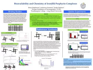
D- Iron Homeostasis, Heme and Porphyrin Synthesis and Metabolism. Introduction.

E N D
D-Iron Homeostasis, Heme and Porphyrin Synthesis and Metabolism
Introduction • Iron serves numerous important functions in the body relating to the metabolism of oxygen, not the least of which is its role in hemoglobin transport of oxygen. Within the body iron exist in two oxidation states: ferrous (Fe2+) or ferric (Fe3+). Because iron has an affinity electronegative atoms such as oxygen, nitrogen and sulfur, these atoms are found at the heart of the iron-binding centers of macromolecules. • Under conditions of neutral or alkaline pH, iron is found in the Fe3+ state and at acidic pH the Fe2+ state is favored. When in the Fe3+ state, iron will form large complexes with anions, water and peroxides. These large complexes have poor solubility and upon their aggregation lead to pathological consequences. In addition, iron can bind to and interfere with the structure and function of various macromolecules. For this reason the body must protect itself against the deleterious effects of iron. This is the role served by numerous iron-binding proteins (see below).
Aside from its importance as the prosthetic group of hemoglobin and a small number of enzymes (e.g., redox cytochromes and the P450 class of detoxifying cytochromes), heme is important because a number of genetic disease states are associated with deficiencies of the enzymes used in its biosynthesis. Some of these disorders are readily diagnosed because they cause δ-aminolevulinic acid, (ALA) and other abnormally colored heme intermediates to appear in the circulation, the urine, and in other tissues such as teeth and bones. Some disorders of heme biosynthesis are more insidious such as the various porphyrias, a list of which can be found below and in the Inborn Errors Page.
Iron Metabolism • Iron is associated with proteins either by incorporation into protoporphyrin IX of by binding to other ligands. When the ferrous form of iron and protoporphyrin IX are complexed the structure is referred to as heme. There are a number of heme containing proteins involved in the transport of oxygen (hemoglobin), oxygen storage (myoglobin) and enzyme catalysis such as nitric oxide synthase (NOS) and prostaglandin synthase (cyclooxygenase). A number of non-heme iron containing proteins are also known such as the iron-sulfur proteins of oxidative phosphorylation and the iron transport and storage proteins, transferrin and ferritin, respectively. • Iron consumed in the diet is either free iron or heme iron. Free iron in the intestines is reduced from the ferric (Fe3+) to the ferrous (Fe2+) state on the luminal surface of intestinal enterocytes and transported into the cells through the action of the divalent metal transporter, DMT1. Intestinal uptake of heme iron occurs through the interaction of dietary heme with the heme carrier protein (HCP1).
The iron in the heme is then released within the enterocytes via the action the heme catabolizing enzyme heme oxygenase (see below). The iron can be stored within intestinal enterocytes bound to ferritin. Iron is transported across the basolateral membrane of intestinal enterocytes into the circulation, through the action of the transport protein ferroportin (also called IREG1=iron-regulated gene 1). Associated with ferroportin is the enzyme hephaestin (a copper-containing ferroxidase with homology to ceruloplasmin) which oxidizes the ferrous form back to the ferric form. Once in the circulation, ferric form of iron is bound to transferrin and passes through the portal circulation of the liver. The liver is the major storage site for iron. The major site of iron utilization is the bone marrow where it is used in heme synthesis.
Dietary iron in the form of non-heme iron or heme iron is absorbed in the duodenum. Non-heme iron occurs primarily in the ferric state in the gut and is reduced to the ferrous state through the action of ferrireductases. In the duodenum this reduction is carried out primarily by duodenal cytochrome b (DCYTB). There are additional intestinal brush border ferrireductases since it has been shown in mice that loss of DCYTB does not impair iron absorption. Ferrous iron is then taken up by duodenal enterocytes through the action of divalent metal transporter 1 (DMT1). DMT1 is a member of the solute carrier protein family and is thus, also known as SLC11A2. Heme iron is taken up through the action of heme carrier protein 1 (HCP1). Once in the enterocyte heme is degraded through the action of heme oxygenase releasing the ferrous iron. Ferrous iron can be stored in the enterocyte bound to ferritin or released to the circulation through the action of ferroportin (also called IREG1). Iron is transported in the blood bound to transferrin but does so only in the ferric state so during the transport through ferroportin, ferrous iron is oxidized by the ferroxidase identified as hephaestin.
Transferrin, made in the liver, is the serum protein responsible for the transport of iron. Although several metals can bind to transferrin, the highest affinity is for the ferric (Fe3+) form of iron. The ferrous form of iron does not bind to transferrin. Transferrin can bind two moles of iron. Cells take up the transported iron through interaction of transferrin with cell-surface receptors. Internalization of the iron-transferrin-receptor complexes is initiated following receptor phosphorylation by PKC. Following internalization, the iron is released due to the acidic nature of the lysosomes. The transferrin receptor is then recycled back to the cell surface.
The main cellular site of iron storage is the liver, specifically in hepatocytes. Iron bound to transferrin is taken up from the blood by hepatocytes due to the binding of transferrin to the transferrin receptor. Free iron (called non-transferrin bound iron, NTBI) in the plasma can also be absorbed by hepatocytes via the action of DMT1. However, the ferric form predominates in the blood and must first be reduced by ferrireductases prior to DMT1 transport. Upon binding transferrin, the transferrin receptor is internalized via receptor-mediated endocytosis. The acidic environment of the endosome results in the release of ferric iron from transferrin. The ferric iron is reduced in the endosome to the ferrous form via the action of an endosomal ferrireductase, most likely STEAP3 (six-transmembrane epithelial antigen of prostate protein 3). The ferrous iron is transported out of the endosome via DMT1 action and can then be stored in the hepatocyte bound to ferritin as in intestinal enterocytes. The transferrin-transferrin receptor complexes are recycled back to the surface of the hepatocyte and the transferrin is released to the blood where it can bind more ferric iron in the circulation. Ferrous iron is released from hepatocytes to the circulation through the action of ferroportin. When in the circulation ferrous iron is oxidized to the ferric form by the plasma ferroxidase known as ceruloplasmin. The ferric iron can then be bound by treansferrin and delivered to other tissues of the body.
Ferritin is the major protein used for intracellular storage of iron. Ferritin without bound iron is referred to as apo-ferritin. Apo-ferritin is a large polymer of 24 polypeptide subunits. This multimeric structure of apo-ferritin is able to bind up to 2,000 iron atoms in the form of ferric-phosphate. The majority of intracellularly stored iron is found in the liver, skeletal muscle and reticuloendothelial cells. If the storage capacity of the ferritin is exceeded, iron will deposit adjacent to the ferritin-iron complexes in the cell. Histologically these amorphous iron deposits are referred to as hemosiderin. Hemosiderin is composed of ferritin, denatured ferritin, and other materials and its molecular structure is poorly defined. The iron present in hemosiderin is not readily available to the cell and thus, cannot supply iron to the cell when it is needed. Hemosiderin is found most frequently in macrophages and is most abundant following hemorrhagic events. • In humans approximately 70% of total body iron is found in hemoglobin. Because of storage and recycling very little (1-2mg) iron will need to be replaced from the diet on a daily basis. Any excess dietary iron is not absorbed or is stored in intestinal enterocytes. Refinement in our understanding of the regulation of iron absorption, recycling and release from intracellular stores has expanded recently with the discovery of the actions of the hepatic iron regulatory protein hepcidin.
Hepcidin was initially described as a 25 amino acid peptide resembling cysteine-rich antimicrobial peptides. Recent evidence has demonstrated that hepcidin functions by inhibiting the presentation of one or more of the iron transporters (e.g. DMT1 and Ireg1) in intestinal membranes. With a high iron diet the level of hepcidin mRNA increases and conversely its levels decrease when dietary iron is low. This is occurring simultaneous to reciprocal changes in the levels of the transporters. Whether hepcidin regulates gene expression or the localization of the intestinal iron transporters is not yet fully understood. • The regulation of iron utilization in the body is primarily controlled via iron-mediated regulation of mRNA translation. The description of this process can be found in the Protein Synthesis page. Both the transferrin receptor and the ferritin mRNAs contain stem-loop structures termed iron responsive elements, IREs. These IREs are recognized by an iron-binding protein containing an iron-sulfur center similar to that of the TCA cycle enzyme aconitase. Other IRE containing mRNAs include those encoding the erythrocyte protoporphyrin synthesis enzyme, ALA synthase (see below), mitochondrial aconitase and Ireg1 (ferroportin).
Clinical Aspects of Abnormal Iron Metabolism • Iron can bind to and form complexes with numerous macromolecules, the consequences of which can be a disruption in normal activities of the affected complexes. Excess intracellular iron results in formation and deposition of hemosiderin which can lead to cellular dysfunction and damage. Thus, the consequences of excess iron intake and storage can have profound consequences. However, one must also consider that a reduction in iron intake can also lead to untoward consequences. Most notably, a reduced iron level negatively affects the function of oxygen transport in red blood cells. Defects in iron metabolism can result from impaired intestinal absorption, excess loss of heme iron due to bleeding as well as to mutations in the iron response elements of iron regulated mRNAs. • Hemochromatosis is defined as a disorder in iron metabolism that is characterized by excess iron absorption, saturation of iron-binding proteins and deposition of hemosiderin in the tissues. The primary affected tissues are the liver pancreas and skin. Iron deposition in the liver leads to cirrhosis and in the pancreas causes diabetes. The excess iron deposition leads to bronze pigmentation of the organs and skin. In fact, the bronze skin pigmentation seen in hemochromatosis, coupled with the resultant diabetes lead to the designation of this condition as bronze diabetes.
The primary cause of hemochromatosis is the inheritance of an autosomal recessive allele. The locus causing hemochromatosis has been designated the HFE1 and is a major histocompatibility complex (MHC) class-1 gene located on chromosome 6. The gene encodes an α chain protein with three immunoglobulin-like domains. This α chain protein associates with β2-microglobulin. Normal HFE1 has been shown to form a complex with the transferrin receptor and in so doing is thought to regulate the rate of iron transfer into cells. A mutation in HFE1 will therefore, lead to increased iron uptake and storage. • The majority of hereditary hemochromatosis patients have inherited a mutation in HFE1 that results in the substitution of Cys 282 for a Tyr (C282Y). This mutation causes loss of conformation of one of the immunoglobulin domains in HFE1. Another mutation found in HFE1 causes a change of His 63 to Asp (H63D). • There are several additional causes of iron overload, although none are as common as classic hemochromatosis. There are at least four additional genetic loci, that when defective lead to hemochromatosis. Two of which are juvenile forms identified as type 2A and 2B and two additional forms identified type 3 and type 4. See the hemochromatosis page for more information.
GRACILE syndrome (GRACILE = growth retardation, aminoacidurina, cholestasis, iron overload, lactic acidosis, early death) is a very rare neonatal lethal disorder caused by defects in the BCS1L gene located on chromosome 2q33. BCS1L stands for BCS1-like which is the human homolog of a yeast gene identified as BCS1. BCS1L encodes a member of the AAA family of ATPases that is necessary for the assembly of complex III of oxidative phosphorylation. Defects in BCSL1 are also responsible for Björnstad syndrome. • Iron deficiency anemia is characterized by microcytic (small) and hypochromic (low pigment) red blood cells. Reduced iron intake and/or excess iron excretion results in a decreased globin protein content in red blood cells as a consequence of the heme control of globin synthesis (see the Protein Synthesis page for details). • The most common causes of iron deficient anemia are excess menstrual flow or gastrointestinal (GI) bleeding. Causes of GI bleeding can include the use of medications that lead to ulceration or erosion of the gastric mucosa, peptic ulcer disease, gastric tumors, hiatal hernia or the gastritis associated with chronic alcohol consumption. • Treatment of iron deficiency anemia is to first determine the cause and source of the excess bleeding. Oral administration of ferrous sulfate is commonly used to supplement the iron loss, however, intravenous iron therapy may be called for in some cases. Severe iron deficiency anemia may necessitate transfusion with packed red blood cells.
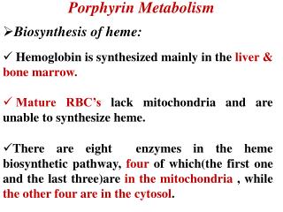
Synthesis of Porphobilinogen and Heme • The first reaction in heme biosynthesis takes place in the mitochondrion and involves the condensation of one glycine and one succinylCoA by the pyridoxal phosphate-containing enzyme, δ-aminolevulinic acid synthase (ALAS). Delta-aminolevulinic acid (ALA) is also called 5-aminolevulinic acid. This reaction is both the rate-limiting reaction of heme biosynthesis, and the most highly regulated reaction (see Regulation below). • There are two forms of ALAS. ALAS1 is considered a house-keeping gene and is expressed in all cells. ALAS2 is an erythroid-specific form of the enzyme and is expressed only in fetal liver and adult bone marrow. The ALAS1 gene is located on chromosome 3, whereas the ALAS2 gene is located on the X chromosome. Deficiencies in ALAS2 result a disorder called X-linked sideroblastic anemia, XLSA. Sideroblasts are erythroblasts with non-heme iron-containing organelles, called siderosomes. XLSA has also been called congenital sideroblastic anemia, hereditary sideroblastic anemia, hereditary iron-loading anemia, X-linked hypochromic anemia, hereditary hypochromic anemia, and hereditary anemia.
Following synthesis mitochondrial ALA is transported to the cytosol, where ALA dehydratase (also called porphobilinogen synthase) dimerizes two molecules of ALA to produce the pyrrole ring compound porphobilinogen. The next step in the pathway involves the head-to-tail condensation of four molecules of porphobilinogen to produce the linear tetrapyrrole intermediate, hydroxymethylbilane. The enzyme for this condensation is porphobilinogen deaminase (PBG deaminase). This enzyme is also called hydroxymethylbilane synthase or uroporphyrinogen I synthase. Hydroxymethylbilane has two main fates. The most important is regulated, enzymatic conversion to uroporphyrinogen III, the next intermediate on the path to heme. This step is mediated by a holoenzyme comprised of uroporphyrinogen synthase plus a protein known as uroporphyrinogen III cosynthase. Hydroxymethylbilane can also non-enzymatically cyclize forming uroporphyrinogen I. • In the Figure below you can place your mouse over intermediate names to see their structures. Clicking on enzyme names will take you to a descriptive page of the porphyria resulting from deficiency in that enzyme.
Pathway of Heme Biosynthesis: PBG = porfobilinógeno (porphobilinogen); ALA = ácido δ-aminolevulínico (δ-aminolevulinc acid)
In the cytosol, the acetate substituents of uroporphyrinogen (normal uroporphyrinogen III or abnormal uroporphyrinogen I) are all decarboxylated by the enzyme uroporphyrinogen decarboxylase. The resultant products have methyl groups in place of acetate and are known as coproporphyrinogens, with coproporphyrinogen III being the important normal intermediate in heme synthesis. • Coproporphyrinogen III is transported to the interior of the mitochondrion, where 2 propionate residues are decarboxylated, yielding vinyl substituents on the 2 pyrrole rings. The colorless product is protoporphyrinogen IX. In the mitochondrion, protoporphyrinogen IX is converted to protoporphyrin IX (structure shown below) by protoporphyrinogen IX oxidase. The oxidase reaction requires molecular oxygen and results in the loss of 6 protons and 6 electrons, yielding a completely conjugated ring system, which is responsible for the characteristic red color to hemes. The final reaction in heme synthesis also takes place in the mitochondrion and involves the insertion of the iron atom into the ring system generating heme b. The enzyme catalyzing this reaction is known as ferrochelatase.
The enzymes ferrochelatase, ALA synthase and ALA dehydratase (a sulfhydryl containing enzyme) are highly sensitive to inhibition by heavy metal poisoning. Indeed, a characteristic of lead poisoning is an increase in ALA in the circulation in the absence of an increase in porphobilinogen. • In addition to the heme b found in hemoglobin, there are three different forms of heme found in cytochromes such as those involved in the process of oxidative phosphorylation. Cytochromes of the c type contain a modified iron protoporphyrin IX known as heme c. In heme c the 2 vinyl (C=C) side chains are covalently bonded to cysteine sulfhydryl residues of the apoprotein. Only cytochromes of the c type contain covalently bound heme. Heme a is also a modified iron protoporphyrin IX. Heme a is found in cytochromes of the a type and in the chlorophyll of green plants
Regulation of Heme Biosynthesis • Although heme is synthesized in virtually all tissues, the principal sites of synthesis are erythroid cells (≈85%) and hepatocytes (accounting for nearly all the rest of heme synthesis). The differences in these two tissues and their needs for heme result in quite different mechanisms for regulation of heme biosynthesis. • In hepatocytes, heme is required for incorporation into the cytochromes, in particular, the P450 class of cytochromes that are important for detoxification. In addition numerous cytochromes of the oxidative-phosphorylation pathway contain heme. The rate-limiting step in hepatic heme biosynthesis occurs at the ALA synthase catalyzed step, which is the committed step in heme synthesis. The Fe3+ oxidation product of heme is termed hemin. Hemin acts as a feed-back inhibitor on ALA synthase. Hemin also inhibits transport of ALA synthase from the cytosol (its' site of synthesis) into the mitochondria (its' site of action) as well as represses synthesis of the enzyme.
In erythroid cells all of the heme is synthesized for incorporation into hemoglobin and occurs only upon differentiation when synthesis of hemoglobin proceeds. When red cells mature both heme and hemoglobin synthesis ceases. The heme (and hemoglobin) must, therefore, survive for the life of the erythrocyte (normally this is 120 days). In reticulocytes (immature erythrocytes) heme stimulates protein synthesis. The mechanism of this mode of heme-mediate regulation of protein synthesis is described in the Protein Synthesis section. Additionally, control of heme biosynthesis in erythrocytes occurs at numerous sites other than at the level of ALA synthase. Control has been shown to be exerted on ferrochelatase, the enzyme responsible for iron insertion into protoporphyrin IX, and on porphobilinogen deaminase.
Heme Metabolism • The largest repository of heme in the human body is in red blood cells, which have a life span of about 120 days. There is thus a turnover of about 6 g/day of hemoglobin, which presents 2 problems. First, the porphyrin ring is hydrophobic and must be solubilized to be excreted. Second, iron must be conserved for new heme synthesis. • Normally, senescent red blood cells and heme from other sources are engulfed by cells of the reticuloendothelial system. The globin is recycled or converted into amino acids, which in turn are recycled or catabolized as required. Heme is oxidized, with the heme ring being opened by the endoplasmic reticulum enzyme, heme oxygenase. The oxidation step requires heme as a substrate, and any hemin (Fe3+) is reduced to heme (Fe2+) prior to oxidation by heme oxygenase.
The oxidation occurs on a specific carbon producing the linear tetrapyrrole biliverdin, ferric iron (Fe3+), and carbon monoxide (CO). This is the only reaction in the body that is known to produce CO. Most of the CO is excreted through the lungs, with the result that the CO content of expired air is a direct measure of the activity of heme oxygenase in an individual. • In the next reaction a second bridging methylene (between rings III and IV) is reduced by biliverdin reductase, producing bilirubin. Bilirubin is significantly less extensively conjugated than biliverdin causing a change in the color of the molecule from blue-green (biliverdin) to yellow-red (bilirubin). The latter catabolic changes in the structure of tetrapyrroles are responsible for the progressive changes in color of a hematoma, or bruise, in which the damaged tissue changes its color from an initial dark blue to a red-yellow and finally to a yellow color before all the pigment is transported out of the affected tissue. Peripherally arising bilirubin is transported to the liver in association with albumin, where the remaining catabolic reactions take place.
Pathway for the degradation of heme to bilirubin. Substituents: M=methyl, P=proprionic, V=vinyl
In hepatocytes, bilirubin-UDP-glucuronyltransferase (bilirubin-UGT) adds 2 equivalents of glucuronic acid to bilirubin to produce the more water soluble, bilirubin diglucuronide derivative. The increased water solubility of the tetrapyrrole facilitates its excretion with the remainder of the bile as the bile pigments. The UDP-glucuronyltransferase (UGT) gene (UGT1A) is located on chromosome 2q37. Several UGT1A enzymes, including bilirubin-UGT (identified as UGT1A1), are encoded by the UGT1A gene complex. The 5' region of the UGT1A complex contains 13 tandemly arrayed first exons, including 4 pseudo exons. These tandemly arrayed exons are identified as 1A1, 1A2, 1A3, etc. Exons 2, 3, 4, and 5 are located in the UGT1A 3' region. All UGT isoforms contain the same C-terminal domain encoded by exons 2 through 5. Each first exon has its own promoter element. The 9 viable first exons are independently spliced to the common exons 2 through 5 to generate 9 UGT1A transcripts with unique 5-prime ends and identical 3-prime ends. The N-terminal region encoded by each unique first exon determines acceptor substrate specificity, while the 246-amino acid C-terminal region encoded by the 4 common exons specifies interactions with the common donor substrate, UDP-glucuronic acid. The bilirubin-UGT isoform (UGT1A1) consists of 533 amino acids.
Bilirubin diglucuronide • In individuals with abnormally high red cell lysis, or liver damage with obstruction of the bile duct, the bilirubin and its precursors accumulate in the circulation; the result is hyperbilirubinemia, the cause of the abnormal yellowish pigmentation of the eyes and tissues known as jaundice. In normal individuals, intestinal bilirubin is acted on by bacteria to produce the final porphyrin products, urobilinogens and urobilins, that are found in the feces. Bilirubin and its catabolic products are collectively known as the bile pigments.
Clinical Aspect of Heme Metabolism • Clinical problems associated with heme metabolism are of two types. Disorders that arise from defects in the enzymes of heme biosynthesis are termed the porphyrias (see Table below and the Porphyrias page) and cause elevations in the serum and urine content of intermediates in heme synthesis. Inherited disorders in bilirubin metabolism lead to hyperbilirubinemia (see the Bilirubinemias page). • Excess circulation and accumulation of bilirubin (hyperbilirubinemia) results in a yellow-orange discoloration of the tissues and is most easily visible as icteric (yellowish) discoloration in the sclera of the eyes. Bilirubin toxicity (bilirubin encephalopathy) can be life threatening in neonates. Bilirubin encephalopathy is characterized by yellow discoloration of the basal ganglia in babies with intense jaundice and was first described over a century ago and the term "kernicterus" was coined to describe these physical changes. Any increase in plasma bilirubin above 20mg/dL is considered dangerous in neonates. However, individual differences in bilirubin sensitivity can result in kernicterus at lower bilirubin levels. Kernicterus occurs in infants with severe unconjugated hyperbilirubinemia and in young adults with high serum levels of unconjugated bilirubin. The latter is the result of inherited deficiencies in the enzyme responsible for bilirubin conjugation to glucuronic acid, bilirubin UDP glucuronyl transferase (bilirubin-UGT).
Bilirubin has been shown to inhibit DNA synthesis, uncouple oxidative phosphorylation, and inhibit ATPase activity in brain mitochondria. Bilirubin also inhibits a variety of different classes of enzymes including dehydrogenases, electron transport proteins, hydrolyases, and enzymes of RNA synthesis, protein synthesis and carbohydrate metabolism. All of these toxic effects of bilirubin are reversed by binding to albumin. In fact, albumin plays a vital role in the disposition of bilirubin in the body by keeping the compound in solution and transporting it from its sites of production (primarily bone marrow and spleen) to its site of excretion which is the liver. • Several inherited disorders in bilirubin metabolism have been identified. Gilbert syndrome and the Crigler-Najjar syndromes result from predominantly unconjugated hyperbilirubinemia. Dubin-Johnson syndrome and Rotor syndrome result from conjugated hyperbilirubinemia. Once conjugated to glucuronate, bilirubin is water soluble, therefore, conjugated hyperbilirubinemias are less severe in their symptomology than are the unconjugated hyperbilirubinemias. • The porphyrias are both inherited and acquired disorders in heme synthesis. These disorders are classified as either erythroid or hepatic, depending upon the principal site of expression of the enzyme defect. Eight different porphyrias have been classified encompassing defects in each of the enzymes of heme synthesis. Defects in hepatic uroporhyrinogen decarboxylase (UROD) results in type I porphyria cutanea tarda (PCT I), whereas deficiencies in the non-hepatic forms of UROD result in type II PCT (PCT II). PCT is the most commonly occurring type of porphyria.
It should be noted that no porphyria has been identified resulting from defects in the house-keeping form of ALAS (ALAS1). The most commonly occurring hepatic porphyria is acute intermittent porphyria, AIP which is caused by a defect in porphobilinogen deaminase, (PBG deaminase). This enzyme is also called hydroxymethylbilane synthase or rarely uroporphyrinogen I synthase. • All of the porphyrias lead to excretion of heme biosynthetic byproducts that turn the urine red and when deposited in the teeth turn them reddish brown. Accumulation of these byproducts in the skin renders it extremely sensitive to sunlight causing ulceration and disfiguring scars. Increased hair growth (hypertrichosis) is also a symptom of the porphryias leading to appearance of fine hairs over the entire face and on the extremities. This latter symptom lends to the description of "werewolf syndrome" in many porphyria patients.